Review
. 2020;110(6):444-476.
doi: 10.1159/000503721. Epub 2019 Sep 27.

Neuroendocrine Neoplasms of the Small Bowel and Pancreas

Affiliations
Review

Neuroendocrine Neoplasms of the Small Bowel and Pancreas

Ashley Kieran Clift et al. Neuroendocrinology. 2020.

Abstract

The traditionally promulgated perspectives of neuroendocrine neoplasms (NEN) as rare, indolent tumours are blunt and have been outdated for the last 2 decades. Clear increments in their incidence over the past decades render them increasingly clinically relevant, and at initial diagnosis many present with nodal and/or distant metastases (notably hepatic). The molecular pathogenesis of these tumours is increasingly yet incompletely understood. Those arising from the small bowel (SB) or pancreas typically occur sporadically; the latter may occur within the context of hereditary tumour predisposition syndromes. NENs can also be associated with endocrinopathy of hormonal hypersecretion. Tangible advances in the development of novel biomarkers, functional imaging modalities and therapy are especially applicable to this sub-set of tumours. The management of SB and pancreatic neuroendocrine tumours (NET) may be challenging, and often comprises a multidisciplinary approach wherein surgical, medical, interventional radiological and radiotherapeutic modalities are implemented. This review provides a comprehensive overview of the epidemiology, pathophysiology, diagnosis and treatment of SB and pancreatic NETs. Moreover, we provide an outlook of the future in these tumour types which will include the development of precision oncology frameworks for individualised therapy, multi-analyte predictive biomarkers, artificial intelligence-derived clinical decision support tools and elucidation of the role of the microbiome in NEN development and clinical behaviour.

Keywords: Neuroendocrine neoplasm; Neuroendocrine tumour; Pancreas; Small intestine.


Conflict of interest statement

Disclosure statement:

M. K. is an employee of Wren Laboratories; I. M. M. is a medical consultant for Wren Laboratories. All other authors declare no competing interests.

Figures

Figure 1.
Figure 1.. Changes over time in NET incidence in the SEER database.
Godwin identified the incidence of NETs to be ~1.3/100,000 (red dot - 1973) in the ERG-TNCS database (pre-SEER). A retrospective analysis from the SEER database is represented as blue dots. Solid dots are mean incidence, error bars correspond to 95% confidence interval. The SEER analysis of 1997 [257] first identified an increased incidence. Thereafter, all subsequent evaluations have demonstrated a steadily increasing incidence (blue dots). This increase is exponential (logarithmic analysis of years: linear regression >0.96) and is consistent with a continuous state-wide assimilation of advances in histopathology, imaging and awareness (HIA). The continued increase in “incidence” suggests that maximal detection levels of NET disease have not been reached. This reflects the failure of HIA advances to have fully permeated the US health care environment. Given the linear slope of the analysis it seems that a likely “real” incidence may not be reached for decades. The disproportionate effect of endoscopic surveillance of the colon and upper GI for cancer and GERD needs to be considered in defining the incidence of life risk significant neoplasia. Data is derived from [5,8,19]
Figure 2.
Figure 2.. Clinical utility of the NETest.
As a diagnostic test (A) for small bowel or pancreatic NET disease, the percentage positive score is 95–100% (n=212) (top left). For lung NETs (n=207), the accuracy is similar (95%). CgA, in comparison is positive in ~30% pancreas, 75% of SI and 45% lung of NETs. Surgical resection (B) reduces the NETest consistent with the tumour removal being the source of the circulating genes. An elevated score (red circles) one month after surgery identifies residual disease and predicts recurrence. Low scores (blue circles) at 6 months indicates complete resection. Elevated NETest scores (C) have a prognostic implication. In a monitored cohort (n=34) over 5 years an elevated NETest occurs 12 months (*) before CT or MRI image-confirmation of disease progression. Monitoring the efficacy of somatostatin analogue (SSA) (D) demonstrates that SSA therapy with disease stabilization exhibits a low NETest. A high NETest on an SSA indicates disease progression. Thus, NETest has utility as a monitoring tool.
Figure 3.
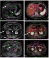
Figure 3.. Benefits of integrating morphological and functional imaging.
Example of the synergistic information deriving from the integration of morphologic (MR, Axial WATER LAVA, 1 min post gadobutrol) and molecular (68Ga-DOTATATE PET/CT, axial fused images) imaging in a patient with G2 neuroendocrine tumor of the ileocecal valve, status post-surgery and referred for restaging of known liver metastases. The MR (A, solid arrow) shows a 0.5 cm lesion in hepatic segment IVA which is not apparent on 68Ga-DOTATATE (B, dotted circle). Other, bigger liver metastases, for example the 1.3 cm lesion identified in segment VI, are concordant on MR (C, solid arrow) and PET (D, solid arrow). In addition, 68Ga-DOTATATE (F, solid arrow) showed intense uptake in a small short-axis node on MRI (E, solid arrow) which did not fulfill the criteria for lymphadenopathy
Figure 4.
Figure 4.. Therapeutic options for small bowel neuroendocrine neoplasms
Treatment algorithm for small bowel neuroendocrine neoplasms, displaying the options available for each sub-type. The ordering of treatments within the same box does not reflect any particular sequencing, rather the options available. NET = neuroendocrine tumour, G = grade, NEC = neuroendocrine carcinoma, SSA = somatostatin analogues, PRRT = peptide receptor radionuclide therapy, IFNa = interferon-alpha, LD + ST = combination of liver-directed and systemic therapies, SIRT = selective internal radiotherapy, TAE = transarterial embolization, TACE = transarterial chemoembolisation. SSAs are not suitable in higher grade NEN due to their de-differentiation and resultant lower expression of somatostatin receptors. Surgery and PRRT have also been utilised in higher grade NEN (G3/NEC) but data are limited to small-size case series.
Figure 5.
Figure 5.. Therapeutic options for pancreatic neuroendocrine neoplasms
Treatment algorithm for pancreatic neuroendocrine neoplasms. The ordering of treatments within the same box does not reflect any particular sequencing, rather the options available. SSAs are not suitable in higher grade NEN due to their de-differentiation and resultant lower expression of somatostatin receptors. Surgery and PRRT have also been utilised in higher grade NEN (G3/NEC), but available data are limited [258]. NET = neuroendocrine tumour, G = grade, NEC = neuroendocrine carcinoma, SSA = somatostatin analogues, PRRT = peptide receptor radionuclide therapy, LD + ST = combination of liver-directed and systemic therapies, SIRT = selective internal radiotherapy, TAE = transarterial embolisation, FOLFIRI = folinic acid & fluorouracil & irinotecan, TACE = transarterial chemoembolisation, STZ/5FU = streptozocin & 5-fluorouracil, CAPTEM = capecitabine & temozolomide, FOLFOX = folinic acid & leucovorin & 5-fluorouracil, CTX = chemotherapy, either streptozotocin/5-fluorouracil (STZ/5-FU) or capecitabine/ temozolomide (CAP/TEM) depending on availability and/or approval MTT, molecular targeted therapy, everolimus or sunitinib
Figure 6.
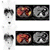
Figure 6.. Response to peptide receptor radionuclide radiotherapy.
Response to PRRT with 177Lu-DOTATATE in a patient affected by a functioning small bowel neuroendocrine tumor with hepatic and nodal metastases, before (upper row, 68Ga-DOTATATE PET/CT: A, MIP image; B, fused axial image; C, axial non-contrast CT) and after (lower row, 68Ga-DOTATATE: B, MIP image; E, fused axial image; F, axial non-contrast CT) treatment. The liver metastases have markedly decreased in extent and tracer avidity at the post-treatment PET/CT, some with increased central photopenia, consistent with central necrosis on CT (A pre-, D, post-treatment, black solid arrow; B, C, pre-, E, F, post-treatment, dotted arrow). The patient also manifested substantial symptomatic improvement (flushing and diarrhoea).
Figure 7.
Figure 7.. Response to multimodal treatment.
Serial 68Ga-SSTR PET/CT MIP images of a 64-year-old woman with well-differentiated, functional pancreatic neuroendocrine neoplasm with liver metastases. The Ki-67 proliferation index was 5% for the primary tumor and 10% for the liver metastasis. Previous treatments were pancreatic tail resection, splenectomy, atypical liver segment resection and open radiofrequency ablation of three liver lesions. 68Ga-DOTATATE PET/CT demonstrates significant somatostatin receptor expression in the hepatic metastases before start of PRRT (A, baseline). The patient was treated with four cycles of 177Lu-DOTATATE (cumulative administered activity 26.4 GBq). Restaging 68Ga-DOTATATE PET/CT at 4 months showed response (B) of the liver metastases, partial remission at 15 and 21 months, respectively (C, D) and complete remission of the disease at 27 months after PRRT (F).

References

    1. Lawrence B, Gustafsson BI, Chan A, Svejda B, Kidd M, Modlin IM: The epidemiology of gastroenteropancreatic neuroendocrine tumors. Endocrinol Metab Clin North Am 2011. Mar;40:1–18, vii. - PubMed
    1. Boyar Cetinkaya R, Aagnes B, Thiis-Evensen E, Tretli S, Bergestuen DS, Hansen S: Trends in Incidence of Neuroendocrine Neoplasms in Norway: A Report of 16,075 Cases from 1993 through 2010. Neuroendocrinology 2017;104:1–10. - PubMed
    1. Frilling A, Clift AK: Therapeutic strategies for neuroendocrine liver metastases. Cancer 2015. Apr 15;121:1172–86. - PubMed
    1. Hauso O, Gustafsson BI, Kidd M, Waldum HL, Drozdov I, Chan AKC, et al.: Neuroendocrine tumor epidemiology: contrasting Norway and North America. Cancer 2008. Nov 15;113:2655–64. - PubMed
    1. Dasari A, Shen C, Halperin D, Zhao B, Zhou S, Xu Y, et al.: Trends in the Incidence, Prevalence, and Survival Outcomes in Patients With Neuroendocrine Tumors in the United States. JAMA Oncol 2017. Oct 1;3:1335. - PMC - PubMed

MeSH terms