. 2019 Sep 25;17(10):548.
doi: 10.3390/md17100548.

Effects of Sulfated Fucans from Laminaria hyperborea Regarding VEGF Secretion, Cell Viability, and Oxidative Stress and Correlation with Molecular Weight

Affiliations

Effects of Sulfated Fucans from Laminaria hyperborea Regarding VEGF Secretion, Cell Viability, and Oxidative Stress and Correlation with Molecular Weight

Philipp Dörschmann et al. Mar Drugs. .

Abstract

Background: Sulfated fucans show interesting effects in the treatment of ocular diseases (e.g., age-related macular degeneration), depending on their chemical structure. Here, we compared three purified sulfated fucans from Laminaria hyperborea (LH) regarding cell viability, oxidative stress protection, and vascular endothelial growth factor (VEGF) secretion in ocular cells.

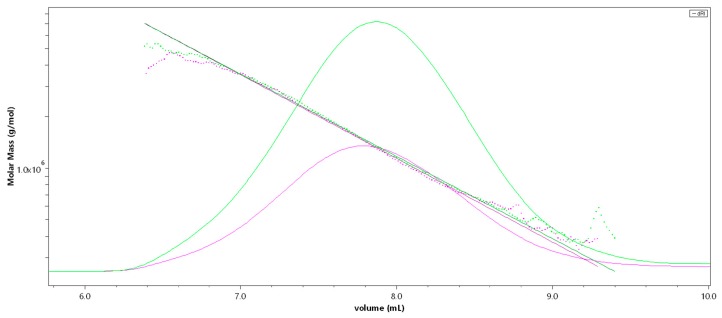
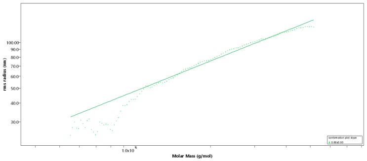
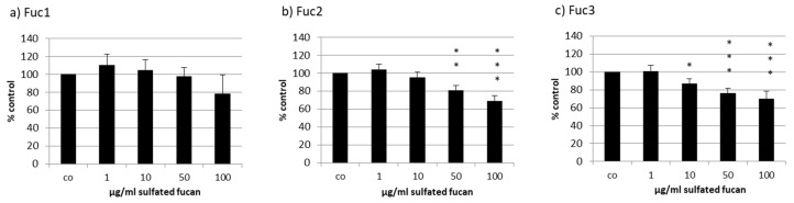
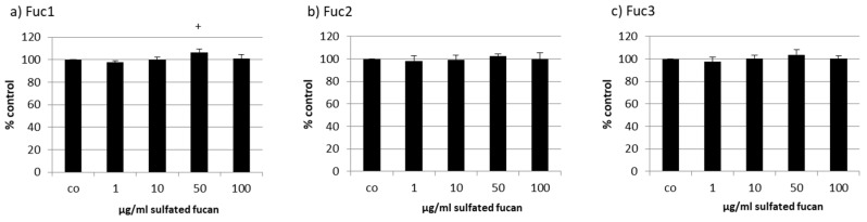
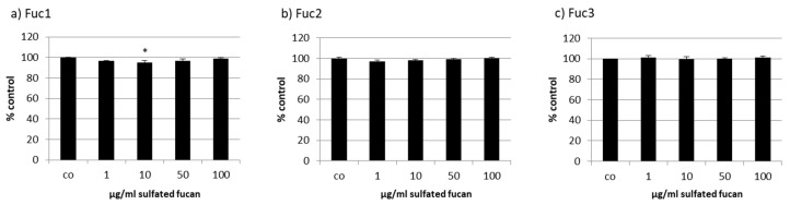
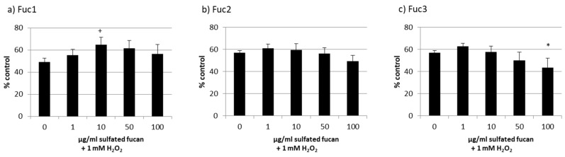
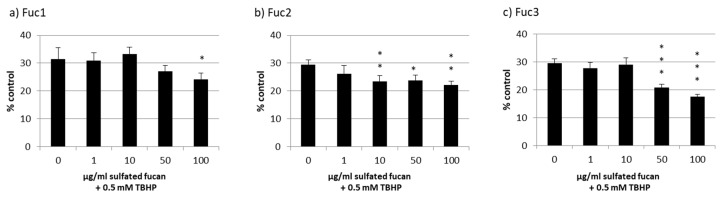
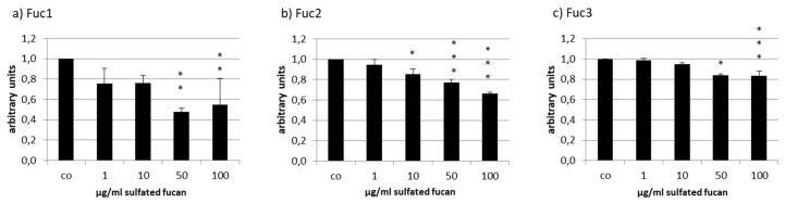
Methods: High-molecular-weight sulfated fucan (Mw = 1548.6 kDa, Fuc1) was extracted with warm water and purified through ultrafiltration. Lower-molecular-weight samples (Mw = 499 kDa, Fuc2; 26.9 kDa, Fuc3) were obtained by mild acid hydrolysis of ultrapurified sulfated fucan and analyzed (SEC-MALS (Size-exclusion chromatography-Multi-Angle Light Scattering), ICP-MS, and GC). Concentrations between 1 and 100 µg/mL were tested. Cell viability was measured after 24 h (uveal melanoma cell line (OMM-1), retinal pigment epithelium (RPE) cell line ARPE-19, primary RPE cells) via MTT/MTS (3-(4,5-dimethylthiazol-2-yl)-2,5-diphenyltetrazolium bromide/3-(4,5-Dimethylthiazol-2-yl)-5-(3-carboxymethoxyphenyl)-2-(4-sulfophenyl)-2H-tetrazolium) assay. Oxidative stress protection was determined after 24 h (OMM-1, ARPE-19). VEGF secretion was analyzed via ELISA after three days (ARPE-19, RPE).

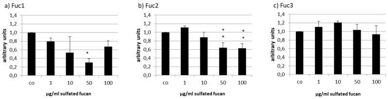
Results: Fuc2 and Fuc3 were antiproliferative for OMM-1, but not for ARPE. Fuc1 protected OMM-1. VEGF secretion was lowered with all fucans except Fuc3 in ARPE-19 and RPE. The results suggest a correlation between molecular weight and biological activity, with efficiency increasing with size.

Conclusion: The LH sulfated fucan Fuc1 showed promising results regarding VEGF inhibition and protection, encouraging further medical research.

Keywords: Laminaria hyperborea; VEGF; age-related macular degeneration; brown seaweed extracts; fucan; fucoidan; molecular weight; oxidative stress; proliferation; retinal pigment epithelium.


Conflict of interest statement

The authors declare no conflict of interest.

Figures

Figure 1
Figure 1
SEC-MALS (Size-exclusion chromatography-Multi-Angle Light Scattering) chromatogram of the high-molecular-weight sulfated fucan (Fuc1) giving M [g/mol] versus V [mL]; 50 µL injection (pink), 100 µL injection (green). The molar mass is plotted as a dotted line, the refractive index is displayed as an overlay.
Figure 2
Figure 2
Rms conformation plot of the high-molecular-weight sulfated fucan (Fuc1) giving the rms radius [nm] versus M [g/mol], 100 µL injection. The slope (b) = 0.66 indicates a random coil.
Figure 3
Figure 3
Cell viability was tested in uveal melanoma cell line OMM-1 after treatment with Laminaria hyperborea (LH) sulfated fucans Fuc1 (a), Fuc2 (b), and Fuc3 (c) for 24 h. Cell viability was determined via MTS (3-(4,5-Dimethylthiazol-2-yl)-5-(3-carboxymethoxyphenyl)-2-(4-sulfophenyl)-2H-tetrazolium) assay and is shown as the mean and standard deviation in relation to a 100% control. Significance was evaluated with ANOVA; * p < 0.05, ** p < 0.01, *** p < 0.001 compared to control (n = 4).
Figure 4
Figure 4
Cell viability was tested in retinal pigment epithelium (RPE) cell line ARPE-19 after treatment with LH sulfated fucans Fuc1 (a), Fuc2 (b), and Fuc3 (c) for 24 h. Cell viability was determined via MTS assay and is shown as the mean and standard deviation in relation to a 100% control. Significance was evaluated with ANOVA compared to the control (n = 4); + p < 0.05.
Figure 5
Figure 5
Cell viability was tested in RPE cells after treatment with LH sulfated fucans Fuc1 (a), Fuc2 (b), and Fuc3 (c) for 24 h. Cell viability was determined via MTT (3-(4,5-dimethylthiazol-2-yl)-2,5-diphenyltetrazolium bromide) assay and is shown as the mean and standard deviation in relation to an untreated control (100%). Significance was evaluated with ANOVA compared to the control (n = 3); * p < 0.05.
Figure 6
Figure 6
OMM-1 cell viability after treatment with Fuc1 (a), Fuc2 (b), and Fuc3 (c) and stress insult after 30 min incubation with the sulfated fucans. The oxidative agent was 1 mM H2O2, which reduced cell viability to under 60% in all cases. Viability was tested via MTS assay and is shown as the mean and standard deviation in relation to an untreated control (100%). The 10 µg/mL Fuc1 showed significant protective effects for the cell line. Significance was evaluated via ANOVA; */+ p < 0.05, versus 0 µg/mL sulfated fucan + 1 mM H2O2 (n = 4).
Figure 7
Figure 7
ARPE-19 cell viability after treatment with Fuc1 (a), Fuc2 (b), and Fuc3 (c) and stress insult after 30 min incubation with the sulfated fucans. The oxidative agent was 0.5 mM tert-butyl hydroperoxide (TBHP), which reduced cell viability to nearly 30% in all cases. Viability was tested via MTS assay and is shown as the mean and standard deviation in relation to an untreated control (100%). No sulfated fucan showed a significant protective effect for the RPE cell line. Significance was evaluated with ANOVA; * p < 0.05, ** p < 0.01, *** p < 0.001, versus 0 µg/mL sulfated fucan + 0.5 mM TBHP (n = 4).
Figure 8
Figure 8
Vascular endothelial growth factor (VEGF) secretion of ARPE19 cells after incubation with Fuc1 (a), Fuc2 (b), and Fuc3 (c). VEGF content was analyzed with ELISA and normalized to cell viability. All LH extracts reduced VEGF content with 50 µg/mL Fuc1 as the most efficient. Significance was evaluated with ANOVA; * p < 0.05, ** p < 0.01, *** p < 0.001 compared to the control (n = 4).
Figure 9
Figure 9
VEGF secretion of RPE cells after incubation with Fuc1 (a), Fuc2 (b), and Fuc3 (c). VEGF content was analyzed via ELISA and normalized to the cell viability. Fuc2 and Fuc1 reduced the VEGF content. Significance was evaluated via ANOVA; * p < 0.05, **p < 0.01, compared to the control (n = 3).

References

    1. Deniaud-Bouët E., Hardouin K., Potin P., Kloareg B., Hervé C. A review about brown algal cell walls and fucose-containing sulfated polysaccharides: Cell wall context, biomedical properties and key research challenges. Carbohydr. Polym. 2017;175:395–408. doi: 10.1016/j.carbpol.2017.07.082. - DOI - PubMed
    1. Li B., Lu F., Wei X., Zhao R. Fucoidan: Structure and bioactivity. Molecules. 2008;13:1671–1695. doi: 10.3390/molecules13081671. - DOI - PMC - PubMed
    1. Klettner A. Fucoidan as a Potential Therapeutic for Major Blinding Diseases—A Hypothesis. Mar. Drugs. 2016;14:31. doi: 10.3390/md14020031. - DOI - PMC - PubMed
    1. Dithmer M., Fuchs S., Shi Y., Schmidt H., Richert E., Roider J., Klettner A. Fucoidan reduces secretion and expression of vascular endothelial growth factor in the retinal pigment epithelium and reduces angiogenesis in vitro. PLoS ONE. 2014;9:e89150. doi: 10.1371/journal.pone.0089150. - DOI - PMC - PubMed
    1. Miller J.W. Age-related macular degeneration revisited—piecing the puzzle: The LXIX Edward Jackson memorial lecture. Am. J. Ophthalmol. 2013;155:1–35.e13. doi: 10.1016/j.ajo.2012.10.018. - DOI - PubMed

LinkOut - more resources