. 2019 Sep 25;11(10):1429.
doi: 10.3390/cancers11101429.

The Targeting of RNA Polymerase I Transcription Using CX-5461 in Combination with Radiation Enhances Tumour Cell Killing Effects in Human Solid Cancers

Affiliations

The Targeting of RNA Polymerase I Transcription Using CX-5461 in Combination with Radiation Enhances Tumour Cell Killing Effects in Human Solid Cancers

Mohammed Ismael et al. Cancers (Basel). .

Abstract

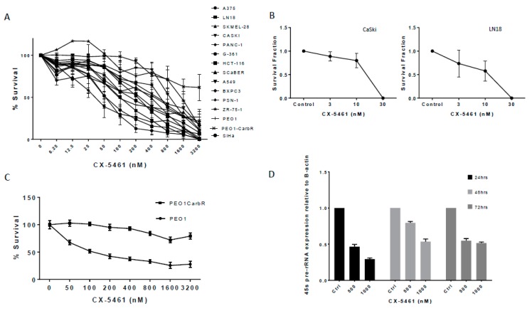
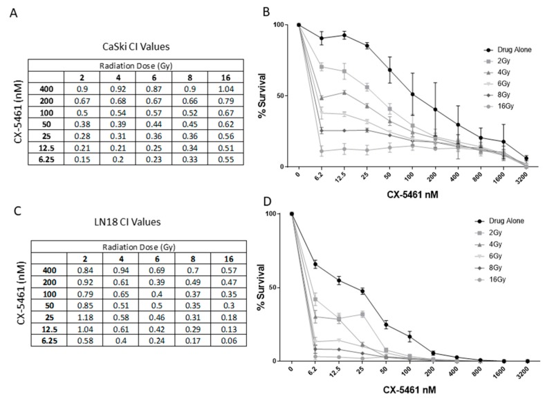
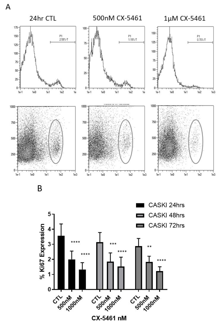
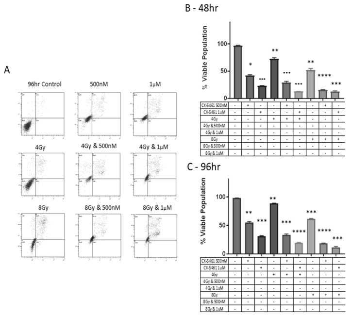
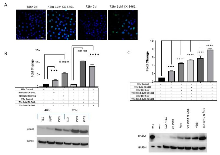
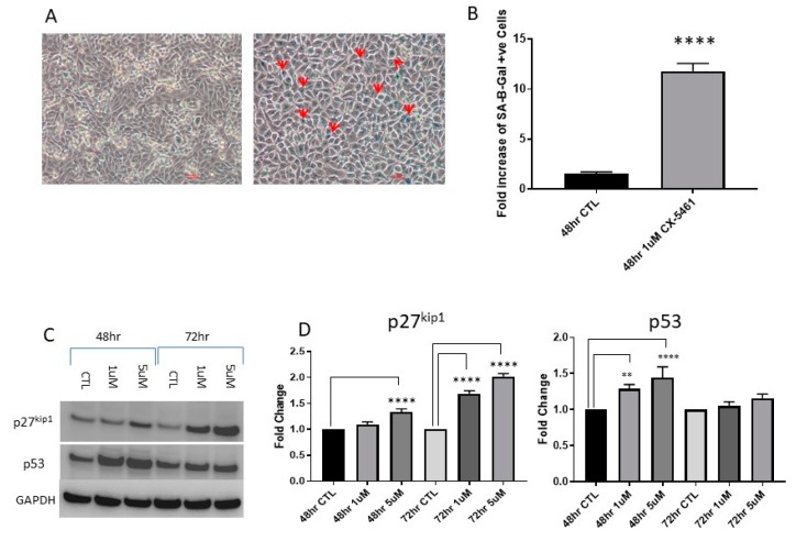
An increased rate of cellular proliferation is a hallmark of cancer and may be accompanied by an increase in ribosome biogenesis and dysregulation in rRNA synthesis. In this regard, CX-5461 has been developed as a novel RNA polymerase I inhibitor and is currently in Phase I/II clinical trials for solid and hematological malignancies. In the present study, interactions between CX-5461 and single-dose X-ray exposure were assessed using isobologram analysis using MTS assay and drug-induced cell death was assessed using flow cytometric, confocal microscopy and Western blot analysis. Combination treatments involving CX-5461 and single-dose X-ray exposure highlighted increased effectiveness compared to individual treatment alone in the CaSki cervical cancer line, with marked synergistic interaction occurring within the low-drug (50 nM) and low-dose radiation range (2-6 Gy). Cell lines challenged with CX-5461 demonstrated the presence of DNA damage, induction of apoptosis, autophagy and senescence alongside high percentages of G2/M cell cycle arrest. In addition, we report preferential sensitivity of ovarian cancer cells with BRCA2 mutation to this novel agent. Taken together, CX-5461 displayed a broad spectrum of activity in a panel of solid cancer cell lines with IC50 values ranging from 35 nM to >1 µM. The work described herein identifies the synergistic effects of CX-5461 in combination with X-rays in solid cancers and may also aid in the design of clinical trials involving this novel agent.

Keywords: CX-5461; RNA polymerase I targeting; combination studies.


Conflict of interest statement

The authors declare no conflict of interest.

Figures

Figure 1
Figure 1
CX-5461 is a potent inhibitor of RNA Polymerase I in solid cancer cell lines. A panel of fifteen solid cancer cells lines was exposed to CX-5461 over 96 h with cell viability assessment using the MTS assay (A). Each cell line was set up in triplicate on the day of experiments and repeated on three separate occasions. (B) CX-5461 as an anti-proliferative agent. Graphs showing survival fraction values of CaSki (top) and LN18 (bottom) following exposure to CX-5461. Doses of 30 nM were sufficient to prevent the formation of colonies in both cell lines. (C) Cell viability data obtained using the MTS assay in PEO1 (BRCA mutant) and PEO1 carboplatin resistant (BRCA wild-type) cell lines showing the difference in sensitivity towards CX-5461. (D) qPCR evaluation of 45s pre rRNA transcription of CaSki cell line following exposure to CX-5461 over 72 h. cT values were normalised against the housekeeping gene B-actin. Transcription of 45s pre rRNA was found to significantly decrease (p-value < 0.0001) in a dose-dependent manner.
Figure 2
Figure 2
The cytotoxic activity of CX-5461 is enhanced when combined with single-dose X-rays. Combination index values obtained through isobologram analysis for CaSki cell line (A and B) and LN18 cell line (C and D) following combination treatments involving CX-5461 and single-dose X-ray exposure (2, 4, 6, 8 and 16 Gy). CI values < 1 were considered synergistic and CI values > 1 were considered antagonistic.
Figure 3
Figure 3
CX-5461 as an anti-proliferative agent. Flow cytometry histogram analysis of CaSki cells probed for Ki67 expression following exposure to sublethal and lethal doses of CX-5461 over 24 h (A) revealed that Ki67 expression was seen to decrease in a dose-dependent and time dependent manner (B) with a p-value of < 0.0001, highlighting the anti-proliferative effects of CX-5461. ** = p value < 0.01, *** = p value < 0.001 and **** = p value < 0.0001.
Figure 4
Figure 4
Flow cytometry analysis of combination treatments involving CX-5461 and single-dose X-ray exposure and the induction of apoptosis. Cell death was monitored using Annexin/PI assay at over 96 h. Histogram plots indicate that CX-5461 treatment alone significantly reduced viable cell populations below 50% at doses of 500 nM and 1 µM in CaSki cell line. Exposure to CX-5461 was shown to induce late apoptosis as determined by high-level annexin and high-level PI staining (A). When combined with X-rays, the cytotoxic effects of CX-5461 were enhanced when combined with single doses of 4 Gy and 1 µM (p-value < 0.001) and further enhanced when using 8 Gy and 1 µM CX-5461 (p-value < 0.001). The enhanced effect of combining CX-5461 alongside X-rays was seen as early as 48 h post treatment (B) through to 96 h (C). ** = p value < 0.01, *** = p value < 0.001 and **** = p value < 0.0001.
Figure 5
Figure 5
CX-5461 treatment and the induction of autophagy. CaSki cells were treated with 1 µM CX-5461 for 48 h and stained for LC3 puncta using anti-rabbit LC3 (1:1000) and counterstained with DAPI. Rapamycin treatment, a known inducer of autophagy was used as a positive control (A). Distinct visualization of autophagolysosomes puncta could be seen in cells exposed to CX-5461, as well as rapamycin. Comparison of the rations of LC3I:LC3II showed significant increases in LC3II:LC3I ratios, indicating the induction of autophagy (C). To determine if complete autophagic flux is occurring, LC3 expression was monitored via Western blot analysis, alongside p62 expression over 72 h (B). Western data showed complete conversion of LC3I to LC3II accompanied by a reduction in p62 expression over the course of 72 h (B). GAPDH was used as loading control to determine relative protein expression. ** = p value < 0.01, *** = p value < 0.001 and **** = p value < 0.0001. Scale bar: 100 μm.
Figure 6
Figure 6
CX-5461 exposure induced DNA damage. CaSki cells were treated with 1 µM CX-5461 over 72 h and visualized using confocal microscopy, DNA damage was marked by γH2AX staining (A) (anti-rabbit γH2AX 1:1000 (green) DAPI (blue), distinct green foci could be seen in CX-5461-treated samples, indicating the presence of double-stranded DNA breaks. The expression of γH2AX was found to increase in a dose- and time-dependent manner (p-value < 0.0001) (B). CaSki cells treated in combination with CX-5461 and single-dose X-rays also resulted in increased expression of γH2AX, as marked by Western blot analysis (C). Densitometry analysis showed that exposure alone to CX-5461 or radiation induced γH2AX expression but combination of 4 Gy and 1 µM CX-5461 and 8 Gy and 1 µM CX-5461 significantly increased γH2AX expression compared to either agent alone (p-value < 0.0001) (C). *** = p value < 0.001 and **** = p value < 0.0001. Scale bar 100 μm.
Figure 7
Figure 7
CX-5461 exposure induced cellular senescence. A375 cells where treated with 1 µM CX-5461 and stained for the presence of B-galactosidase following 48 h CX-461 treatment and imaged using brightfield microscopy (A). Cells which have entered into a senescent state are marked by red arrows, (magnification 20×). Analysis of blue pixels in treated and untreated samples using ImageJ software showed a significant increase in senescence-associated b-galactosidase-positive cells in CX-5461-treated melanoma cell line (p-value < 0.0001) (B). Western blot analysis of cell-cycle-associated markers p27kip1 and p53 (C), p53 induction could be seen as early as 48 h post CX-5461 treatment (p-value < 0.0001) in CaSki cell line and strong p27 induction could be observed at 72 h post CX-5461 treatment (D). ** = p value < 0.01 and **** = p value < 0.0001.
Figure 8
Figure 8
Cell cycle analysis of CaSki cells exposed to CX-5461 alone, radiation alone and in combination for 48 h showed that treatment with CX-5461 alone for 48 h induced strong G2/M arrest compared to radiation-alone treatment, combined treatment resulted in the abolition of the cell cycle, as shown by propidium iodide staining using flow cytometry (A). (48 h CaSki control cell cycle values: G0-6%, G1-51%, S-15% G2M-28%. The 1 µM CX-5461 cell cycle values: G0-3%, G1-41%, S-6%, G2M-49%. 4 Gy-radiation-alone cell cycle values: G0-0.8%, G1-53.2%, S-9.2%, G2M-29.4%. Combined 1 µM CX-5461 and 4 Gy X-rays resulted in complete abolition of G2M blockade). NPM1 silenced cells are more sensitive to RNA Pol I inhibition. (B,C) Flow cytometry (annexin assay) showed that NPM1 silenced cells were found to be more sensitive towards CX-5461 treatment compared to non-coding treated CaSki cells (p-value < 0.0001). CaSki cells were transiently silenced for nucleophosmin (NPM1). Knock down was confirmed through Western blot analysis (D). ** = p value < 0.01 and **** = p value < 0.0001.

Similar articles

Cited by

References

    1. Boisvert F.M., van Koningsbruggen S., Navascués J., Lamond A.I. The multifunctional nucleolus. Nat. Rev. Mol. Cell Biol. 2007;8:574–585. doi: 10.1038/nrm2184. - DOI - PubMed
    1. Drygin D., Rice W.G., Grummt I. The RNA Polymerase I transcription machinery—an emerging target for the treatment of cancer. Annu. Rev. Pharmacol. Toxicol. 2010;50:131–156. doi: 10.1146/annurev.pharmtox.010909.105844. - DOI - PubMed
    1. Villicaña C., Cruz G., Zurita M. The basal transcription machinery as a target for cancer therapy. Cancer Cell Int. 2014;14:18. doi: 10.1186/1475-2867-14-18. - DOI - PMC - PubMed
    1. Williamson D., Lu Y.J., Fang C., Pritchard-Jones K., Shipley J. Nascent pre-rRNA overexpression correlates with an adverse prognosis in alveolar rhabdomyosarcoma. Genes Chromosomes Cancer. 2006;45:839–845. doi: 10.1002/gcc.20347. - DOI - PubMed
    1. Derenzini M., Trere D., Pession A., Govoni M., Sirri V., Chieco P. Nucleolar size indicates the rapidity of cell proliferation in cancer tissues. J. Pathol. 2000;191:181–186. doi: 10.1002/(SICI)1096-9896(200006)191:2<181::AID-PATH607>3.0.CO;2-V. - DOI - PubMed

LinkOut - more resources