. 2019 Nov 15;294(46):17371-17382.
doi: 10.1074/jbc.RA119.010072. Epub 2019 Sep 26.

Mutations at hypothetical binding site 2 in insulin and insulin-like growth factors 1 and 2 result in receptor- and hormone-specific responses

Affiliations

Mutations at hypothetical binding site 2 in insulin and insulin-like growth factors 1 and 2 result in receptor- and hormone-specific responses

Kateřina Macháčková et al. J Biol Chem. .

Abstract

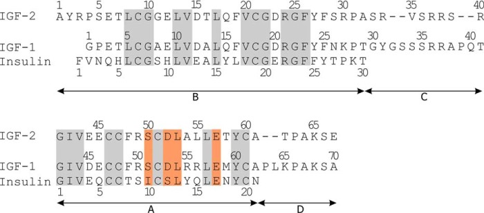
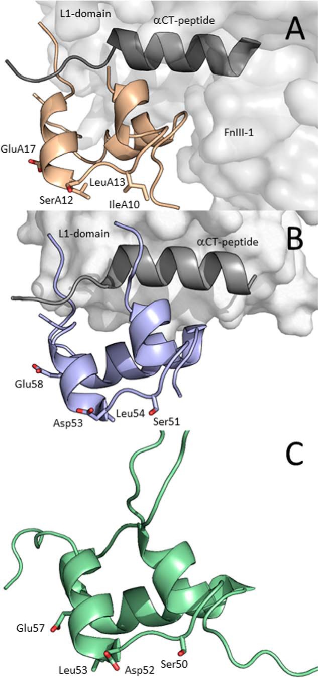
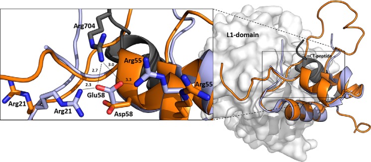
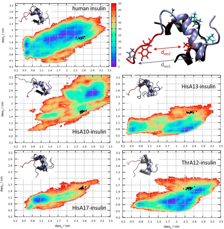
Information on how insulin and insulin-like growth factors 1 and 2 (IGF-1 and -2) activate insulin receptors (IR-A and -B) and the IGF-1 receptor (IGF-1R) is crucial for understanding the difference in the biological activities of these peptide hormones. Cryo-EM studies have revealed that insulin uses its binding sites 1 and 2 to interact with IR-A and have identified several critical residues in binding site 2. However, mutagenesis studies suggest that Ile-A10, Ser-A12, Leu-A13, and Glu-A17 also belong to insulin's site 2. Here, to resolve this discrepancy, we mutated these insulin residues and the equivalent residues in IGFs. Our findings revealed that equivalent mutations in the hormones can result in differential biological effects and that these effects can be receptor-specific. We noted that the insulin positions A10 and A17 are important for its binding to IR-A and IR-B and IGF-1R and that A13 is important only for IR-A and IR-B binding. The IGF-1/IGF-2 positions 51/50 and 54/53 did not appear to play critical roles in receptor binding, but mutations at IGF-1 position 58 and IGF-2 position 57 affected the binding. We propose that IGF-1 Glu-58 interacts with IGF-1R Arg-704 and belongs to IGF-1 site 1, a finding supported by the NMR structure of the less active Asp-58-IGF-1 variant. Computational analyses indicated that the aforementioned mutations can affect internal insulin dynamics and inhibit adoption of a receptor-bound conformation, important for binding to receptor site 1. We provide a molecular model and alternative hypotheses for how the mutated insulin residues affect activity.

Keywords: NMR structure; complex; hormone analog; insulin; insulin-like growth factor (IGF); molecular dynamics; mutagenesis; peptide hormone; receptor autophosphorylation; receptor binding; receptor tyrosine kinase; structural biology; structure-function.


Conflict of interest statement

The authors declare that they have no conflicts of interest with the contents of this article

Figures

Figure 1.
Figure 1.
Primary sequences of human insulin, IGF-1, and IGF-2. The residues mutated in this study are highlighted with an orange background, and homologous residues are highlighted with a gray background.
Figure 2.
Figure 2.
Receptor-bound structures of insulin and IGF-1 and NMR structure of IGF-2. A, cryo-EM structure of IR-A–bound insulin (PDB code 6HN5 from Ref. , in light brown). Receptor site 1′ is represented by the L1 domain (light gray), and αCT peptide (dark gray) and receptor site 2′ are represented by the FnIII-1 domain (light gray). B, crystal structure of IGF-1 (PDB code 5U8Q from Ref. , in violet) bound to L1 domain (in light gray) and αCT (in dark gray) representing site 1′ of IGF-1R. C, NMR structure of human IGF-2 (PDB code 5L3L from Ref. , in green). The side chains of residues modified in this study are shown as sticks and are numbered.
Figure 3.
Figure 3.
An overlay of IGF-1R–bound human IGF-1 with Asp-58–IGF-1. Human IGF-1 is in light blue (PDB code 5U8Q from Ref. 19), and a representative (lowest energy) NMR structure of Asp-58–IGF-1 is in orange (PDB code 6RVA). The receptor site 1′ is represented by L1 domain (in gray) and α-CT peptide (in black). The enlarged window on the left shows side chains of hormones' Glu-58, Asp-58, or Arg-704 (from α-CT) and two other IGF-1 arginines (Arg-21 and Arg-55) as sticks with nitrogen atoms in blue and oxygen atoms in red. Some possible interactions of Glu-58 and Arg-704, Arg-21, and Arg-55 residues identified in the complex are indicated by dashed lines with distances in Å.
Figure 4.
Figure 4.
Free energy profiles (color scale in kJ/mol) of human insulin and analogs obtained from metadynamics. Black dots represent insulin conformations from molecular dynamics simulations in complex with IR. The B-chain of representative minimal energy conformers of insulin mutants (in black) aligned to the IR-bound conformation of human insulin (ice blue, C terminus in red) are depicted in the insets. The IR-bound conformation of human insulin is shown on the upper right, with residues defining the dwo1 (Gly-B8–Pro-B28) and dwo2 (Val-B12–Tyr-B26) distances, represented as ice blue and red licorice, respectively. The A-chain is in black, with the mutated residues (Ile-A10, Ser-A12, Leu-A13, Glu-A17) shown as licorice and colored by atom type.

References

    1. De Meyts P. (2004) Insulin and its receptor: structure, function and evolution. Bioessays 26, 1351–1362 10.1002/bies.20151 - DOI - PubMed
    1. Le Roith D. (2003) The insulin-like growth factor system. Exp. Diabesity Res. 4, 205–212 10.1155/EDR.2003.205 - DOI - PMC - PubMed
    1. Ward C. W., and Lawrence M. C. (2012) Similar but different: ligand-induced activation of the insulin and epidermal growth factor receptor families. Curr. Opin. Struct. Biol. 22, 360–366 10.1016/j.sbi.2012.03.014 - DOI - PubMed
    1. Lemmon M. A., and Schlessinger J. (2010) Cell signaling by receptor tyrosine kinases. Cell 141, 1117–1134 10.1016/j.cell.2010.06.011 - DOI - PMC - PubMed
    1. Belfiore A., Malaguarnera R., Vella V., Lawrence M. C., Sciacca L., Frasca F., Morrione A., and Vigneri R. (2017) Insulin receptor isoforms in physiology and disease: An updated view. Endocr. Rev. 38, 379–431 10.1210/er.2017-00073 - DOI - PMC - PubMed

Publication types

MeSH terms

Supplementary concepts