. 2019 Dec;74(12):1131-1139.
doi: 10.1136/thoraxjnl-2018-212430. Epub 2019 Sep 26.

MUC5B variant is associated with visually and quantitatively detected preclinical pulmonary fibrosis

Affiliations

MUC5B variant is associated with visually and quantitatively detected preclinical pulmonary fibrosis

Susan K Mathai et al. Thorax. 2019 Dec.

Abstract

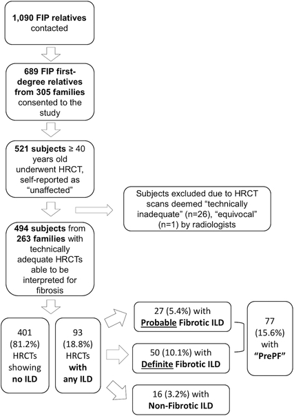
Background: Relatives of patients with familial interstitial pneumonia (FIP) are at increased risk for pulmonary fibrosis. We assessed the prevalence and risk factors for preclinical pulmonary fibrosis (PrePF) in first-degree relatives of patients with FIP and determined the utility of deep learning in detecting PrePF on CT.

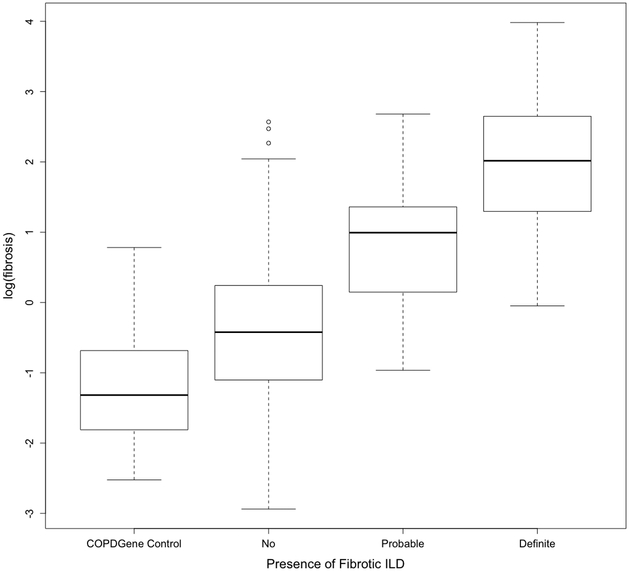
Methods: First-degree relatives of patients with FIP over 40 years of age who believed themselves to be unaffected by pulmonary fibrosis underwent CT scans of the chest. Images were visually reviewed, and a deep learning algorithm was used to quantify lung fibrosis. Genotyping for common idiopathic pulmonary fibrosis risk variants in MUC5B and TERT was performed.

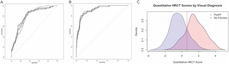
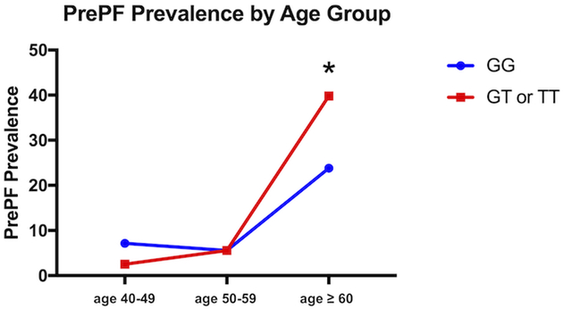
Findings: In 494 relatives of patients with FIP from 263 families of patients with FIP, the prevalence of PrePF on visual CT evaluation was 15.6% (95% CI 12.6 to 19.0). Compared with visual CT evaluation, deep learning quantitative CT analysis had 84% sensitivity (95% CI 0.72 to 0.89) and 86% sensitivity (95% CI 0.83 to 0.89) for discriminating subjects with visual PrePF diagnosis. Subjects with PrePF were older (65.9, SD 10.1 years) than subjects without fibrosis (55.8 SD 8.7 years), more likely to be male (49% vs 37%), more likely to have smoked (44% vs 27%) and more likely to have the MUC5B promoter variant rs35705950 (minor allele frequency 0.29 vs 0.21). MUC5B variant carriers had higher quantitative CT fibrosis scores (mean difference of 0.36%), a difference that remains significant when controlling for age and sex.

Interpretation: PrePF is common in relatives of patients with FIP. Its prevalence increases with age and the presence of a common MUC5B promoter variant. Quantitative CT analysis can detect these imaging abnormalities.

Keywords: Idiopathic pulmonary fibrosis; Imaging/CT MRI etc; Interstitial Fibrosis.


Conflict of interest statement

Competing interests: DAS is the founder and chief scientific officer of Eleven P15, a company focused on the early diagnosis and treatment of pulmonary fibrosis. DAS has an awarded patent (US patent no: 8,673,565) for the treatment and diagnosis of fibrotic lung disease. DAL and SMH have a pending patent (application US20170330320A1) for image analysis; SMH reports a consulting agreement with Boehringer Ingelheim.

Figures

Figure 1.
Figure 1.. Enrollment and Screening Flowchart
Description of enrollment process and results for study subjects.
Figure 2.
Figure 2.. Representative Images from Cohort Subjects
A. High-resolution CT (HRCT) image of the chest from a study subject whose scan was read as normal, without signs of interstitial lung disease or fibrosis. B. HRCT image from subject who was categorized as having “Probable Fibrotic ILD.” C. Representative HRCT image from subject who was characterized as having “Definite Fibrotic ILD.” D. HRCT image from a case of previously diagnosed, established Idiopathic Pulmonary Fibrosis (IPF) in one of the study families.
Figure 3.
Figure 3.. Categorization of Regions of HRCT Images using Quantitative Methodology
Representative axial HRCT images visually assessed as “No Fibrosis” (A), “Probable Fibrotic ILD” (B) and “Definite Fibrotic ILD” (C). Below each is the corresponding quantitative HRCT results for the above scan; regions classified as fibrotic are shown in red. (A) “No Fibrosis” fibrosis extent 0.10% (log(fibrosis score) = −2.30); (B) “Probable Fibrotic ILD” fibrosis extent 12.46% (log(fibrosis score) = 2.52); (C) “Definite Fibrotic ILD” fibrosis extent 24.05% (log(fibrosis score) 3.18).
Figure 4.
Figure 4.. Fibrosis Score by Visual Diagnosis
Boxplots of fibrosis scores based on quantitative HRCT assessment for each visual diagnosis category. Fibrosis score means were significantly different (ANOVA, p<0.0001) across groups defined by visual diagnosis. Comparison of fibrosis score between groups showed significant differences for all individual comparisons (p<0.01 for all).
Figure 5.
Figure 5.. Receiver Operating Characteristic (ROC) Curves for Quantitative Imaging Measures of Fibrosis and PrePF
A. ROC curves for visual diagnosis compared to log %HAA. For this quantitative method, mean AUC was 0.80 (range 0.79–0.81). B. ROC Curves for visual diagnosis compared to fibrosis scores. ROC analysis showed that fibrosis score discriminates subjects with visual diagnosis of PrePF. Average area under the curve (AUC) in five-fold cross validation was 0.92 (range 0.91–0.93) and average accuracy, sensitivity, and specificity in the test partitions were 0.85 (range 0.81–0.88), 0.81 (range 0.71–0.92), and 0.86 (range 0.79–0.90), respectively. Optimal threshold for log fibrosis score was 0.60 (range 0.53 – 0.71), corresponding to 1.8% fibrotic area in examined lung. (C) Density plots of fibrosis scores for visually diagnosed PrePF (pink) and No Fibrosis (blue) scans—the fibrosis score optimal threshold is indicated with the red line (0.60).
Figure 6.
Figure 6.. Prevalence of PrePF in FIP Siblings Cohort by Age and MUC5B Genotype
PrePF prevalence in this FIP siblings cohort increases by age, as shown in this graph. By age > 60 years, the prevalence of PrePF differed significantly based on MUC5B genotype (*p=0.02). Subjects with the variant are depicted by the red line, while those without it are depicted with the blue line.

Comment in

References

    1. Ley B, Collard HR. Epidemiology of Idiopathic Pulmonary Fibrosis. Clin Epidemiol 2013;5:483–92. doi:10.1055/s-2007-1006319 - DOI - PMC - PubMed
    1. Richeldi L, du Bois RM, Raghu G, et al. Efficacy and safety of nintedanib in idiopathic pulmonary fibrosis. N Engl J Med 2014;370:2071–82. doi:10.1056/NEJMoa1402584 - DOI - PubMed
    1. King TE, Bradford WZ, Castro-Bernardini S, et al. A phase 3 trial of pirfenidone in patients with idiopathic pulmonary fibrosis. N Engl J Med 2014;370:2083–92. doi:10.1056/NEJMoa1402582 - DOI - PubMed
    1. Seibold MA, Wise A, Speer M, et al. A common MUC5B promoter polymorphism and pulmonary fibrosis. N Engl J Med 2011;364:1503–12. - PMC - PubMed
    1. Fingerlin TE, Murphy E, Zhang W, et al. Genome-wide association study identifies multiple susceptibility loci for pulmonary fibrosis. Nat Genet 2013;45:613–20. doi:10.1038/ng.2609 - DOI - PMC - PubMed

Publication types

MeSH terms