Review
. 2019 Jul 31;13(7):1-13.
doi: 10.3941/jrcr.v13i7.3607. eCollection 2019 Jul.

Multidisciplinary approach in the management of uterine intravenous leiomyomatosis with intracardiac extension: case report and review of literature

Affiliations
Review

Multidisciplinary approach in the management of uterine intravenous leiomyomatosis with intracardiac extension: case report and review of literature

Gianluca Marrone et al. J Radiol Case Rep. .

Abstract

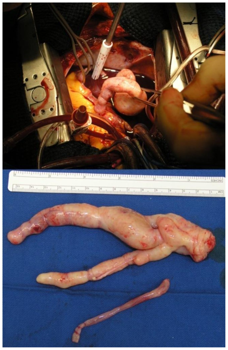
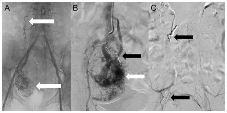
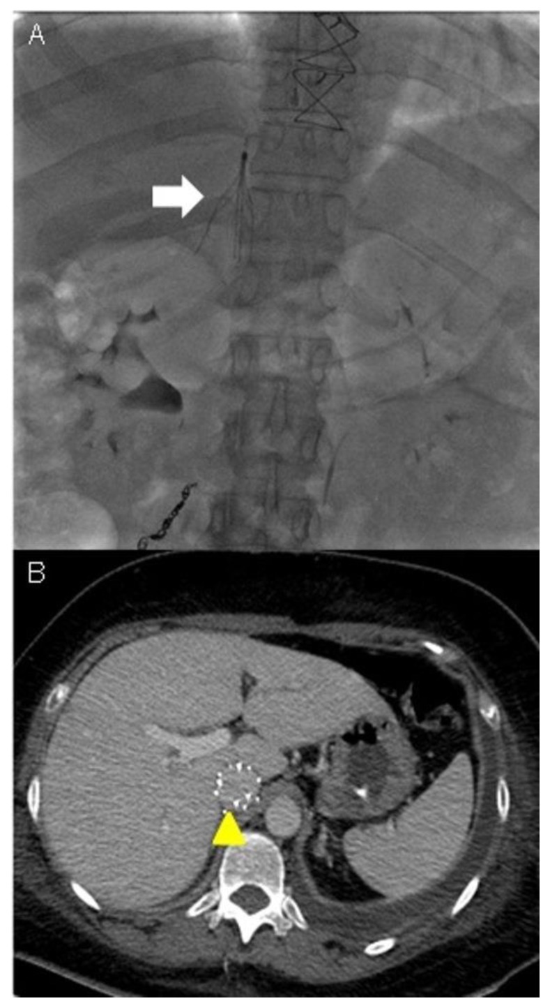
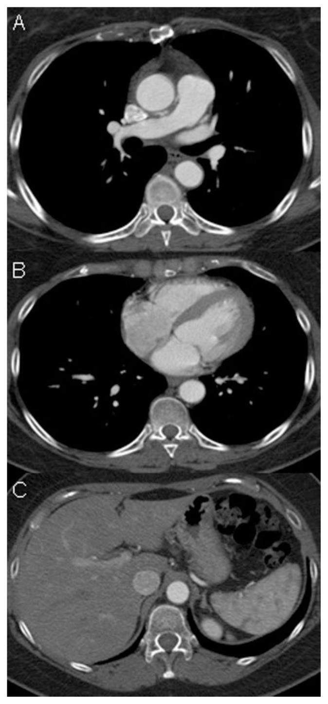
Uterine intravenous leiomyomatosis is an uncommon tumor, usually arising from the uterus, with nodular masses which extend intravascularly over variable distances and may reach the inferior vena cava, right atrium, and pulmonary arteries. Early diagnosis and surgical intervention are crucial as intracardiac leiomyomatosis not only causes cardiac symptoms but may result in pulmonary embolism and sudden death. Complete tumor resection is key in disease management, thus rendering cardiac-extending uterine intravenous leiomyomatosis one of the most challenging conditions for surgical treatment. The use of interventional radiology procedures can facilitate the surgical approach. We report the case of a massive pelvic recurrence of uterine leiomyomatosis with intracardiac extension and pulmonary embolism, analyzing management and surgical outcomes, highlighting the role of interventional radiology during the therapeutic pathway. Nonetheless, there are currently very few data available concerning the use of interventional radiology procedures in the therapeutic strategy of uterine intravenous leiomyomatosis with intracardiac extension.

Keywords: Computed Tomography angiography; Percutaneous embolizazion; Uterine intravenous leiomyomatosis; inferior vena cava filter; intracardiac extension.


Figures

Figure 1
Figure 1
A 48-year-old woman with uterine intravenous leiomyomatosis and intracardiac extension. Technique: x-ray performed in postero-anterior projection, 2mAs, 120kV. GE Connexity. Findings: No significant abnormalities
Figure 2
Figure 2
A 48-year-old woman with uterine intravenous leiomyomatosis and intracardiac extension. Technique: Ultrasound performed on GE LOGIC E9 with a 4C-RS transducer at a frequency of 4.0 MHz. Findings: A) The color-Doppler-ultrasound (CDUS) examination showed a mass in the lower abdomen. B, C) the mass was characterized by intralesional vascularization and a tubular lesion inside the inferior vena cava (IVC), with a triphasic CDUS waveform (see the thin white arrow). CTA is mandatory for further evaluation.
Figure 3
Figure 3
A 48-year-old woman with uterine intravenous leiomyomatosis and intracardiac extension. Technique: Contrast enhanced CT, 249 mA, 120kV, 2.5 mm slice thickness, intravenous contrast: 130 mL of Ultravist (Iopromide). GE LightSpeed 64. Findings: A, B) Axial contrast enhanced CT of the abdomen in the arterial phase demonstrated the presence of a voluminous pelvic mass with significant contrast enhancement due to hypervascularization (see the yellow arrow). C, D, E) Notice the presence of contrast-enhanced large vessels within the IVC (see the thin white arrow).
Figure 4
Figure 4
A 48-year-old woman with uterine intravenous leiomyomatosis and intracardiac extension. Technique: Contrast enhanced CT, 249mA, 120kV, 2.5 mm slice thickness, intravenous contrast: 130 mL of Ultravist (Iopromide). GE LightSpeed 64. Findings: A,B) Coronal and sagittal CT Maximum Intensity Projections (MIP) of the abdomen in the arterial phase showing the pelvic tumor (see the yellow arrowhead) directly extending into the IVC (see the thin white arrows). C,D) 3-D reconstruction from the source data confirmed the origin of the thrombus from a uterine mass invading the right iliac vein and then the IVC.
Figure 5
Figure 5
A 48-year-old woman with uterine intravenous leiomyomatosis and intracardiac extension. Technique: Contrast enhanced CT, 249mA, 120 kV, 2.5 mm slice thickness, intravenous contrast: 130 mL of Ultravist (Iopromide). GE LightSpeed 64. Findings: A, B, C, D) Coronal and sagittal CT projections showing the caval (see the yellow arrow) and cardiac extension of the tumor, up to the right atrium, right ventricle (see the red arrowheads), right ventricle outflow, to the pulmonary trunk (pa).
Figure 6
Figure 6
A 48-year-old woman with uterine intravenous leiomyomatosis and intracardiac extension. Image taken intraoperatively during cardiac surgery for intra-cardiac-intra-caval mass removal. It shows the bizarre morphology of the tumor resembling the intraluminal shape of the IVC in which it runs.
Figure 7
Figure 7
A 48-year-old woman with uterine intravenous leiomyomatosis and intracardiac extension. Technique: Selective embolization of the uterine mass performed using microcatheter Direxion (Direxion HI-FLO™ Torqueable Microcatheters Boston Scientific) and metallic coils. Findings: A, B) The angiogram showed a hypervascular pelvic mass, with anomalous peripheral new vessels formation, invading the IVC (see the white and black arrows). C) A final angiogram showed a drastic reduction of the vascularization (see the black arrows).
Figure 8
Figure 8
A 48-year-old woman with uterine intravenous leiomyomatosis and intracardiac extension. Technique: Contrast enhanced CT, 249mA, 120kV, 2.5 mm slice thickness, intravenous contrast: 130 mL of Ultravist (Iopromide). GE LightSpeed 64. Findings: A, B) An axial contrast enhanced CT of the abdomen performed after percutaneous embolization procedure of the uterine mass showed a relevant reduction in tumor size and vascularization. Note the metallic coils (see the yellow arrows).
Figure 9
Figure 9
A 48-year-old woman with uterine intravenous leiomyomatosis and intracardiac extension. Technique: Under radiologic guidance a catheter was inserted via jugular vein access and advanced to the inferior vena cava in the abdomen. Contrast material was then injected into the vein to assess for proper positioning of the IVC filter. Findings: A) The filter was correctly expanded and attached itself to the walls of the IVC (see the white arrow). B) CTA was used to monitor the results of IVC caval filter. CTA venous phase showed the caval filter in the retrohepatic IVC, of note the anomalous position of the filter was necessary due to the extension of the thrombosis above the renal veins (see the yellow arrowhead).
Figure 10
Figure 10
A 48-year-old woman with uterine intravenous leiomyomatosis and intracardiac extension. Technique: Contrast enhanced CT, 249mA, 120kV, 2.5 mm slice thickness, intravenous contrast: 130 mL of Ultravist (Iopromide). GE LightSpeed 64. Findings: A, B, C) Axial CT images showed complete regression of intravascular and intracardiac leiomyomatosis after the surgical excision. At two-year follow-up the patient showed no evidence of disease recurrence.

References

    1. Fornaris RJ, Rivera M, Jiménez L, Maldonado J. Multimodality Evaluation of Intravenous Leiomyomatosis: A Rare, Benign but Potentially Life-Threatening Tumor. Am J Case Rep. 2015;16:794–800. - PMC - PubMed
    1. Nakai G, Maeda K, Yamamoto K, Yamada T, Hirose Y, Terai Y, Ohmichi M, Katsumata T, Narumi Y. Uterine Intravenous Leiomyomatosis with Cardiac Extension: Radiologic Assessment with Surgical and Pathologic Correlation. Case Rep Obstet Gynecol. 2015;2015 576743. - PMC - PubMed
    1. Gui T, Qian Q, Cao D, Yang J, Peng P, Shen K. Computerized tomography angiography in preoperative assessment of intravenous leiomyomatosis extending to inferior vena cava and heart. BMC Cancer. 2016;16:73. - PMC - PubMed
    1. Xu ZF, Yong F, Chen YY, Pan AZ. Uterine intravenous leiomyomatosis with cardiac extension: Imaging characteristics and literature review. World J Clin Oncol. 2013;4(1):25–28. - PMC - PubMed
    1. Lee S, Kim DK, Narm KS, Cho SH. Pulmonary artery embolization of intravenous leiomyomatosis extending into the right atrium. Korean J Thorac Cardiovasc Surg. 2011;44(3):243–6. - PMC - PubMed

MeSH terms