Review
. 2019 Aug 31;13(8):1-18.
doi: 10.3941/jrcr.v13i8.3633. eCollection 2019 Aug.

Intracranial calcifications on CT: an updated review

Affiliations
Review

Intracranial calcifications on CT: an updated review

Charbel Saade et al. J Radiol Case Rep. .

Abstract

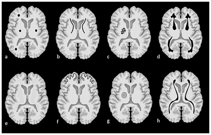
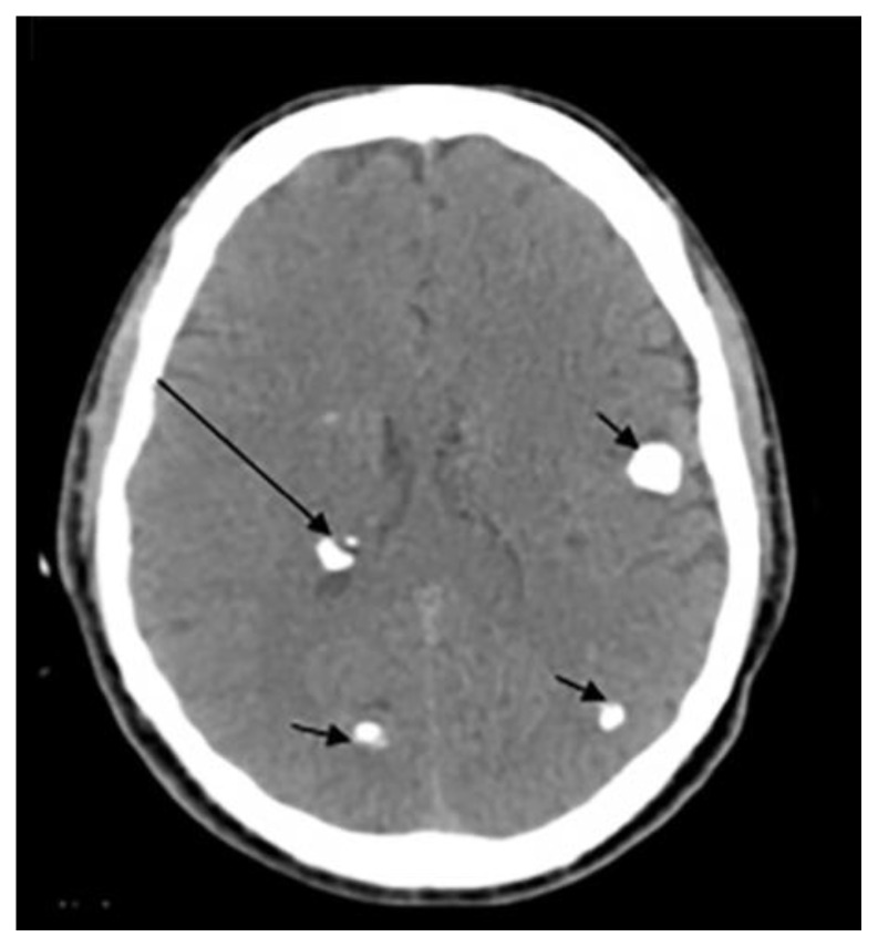
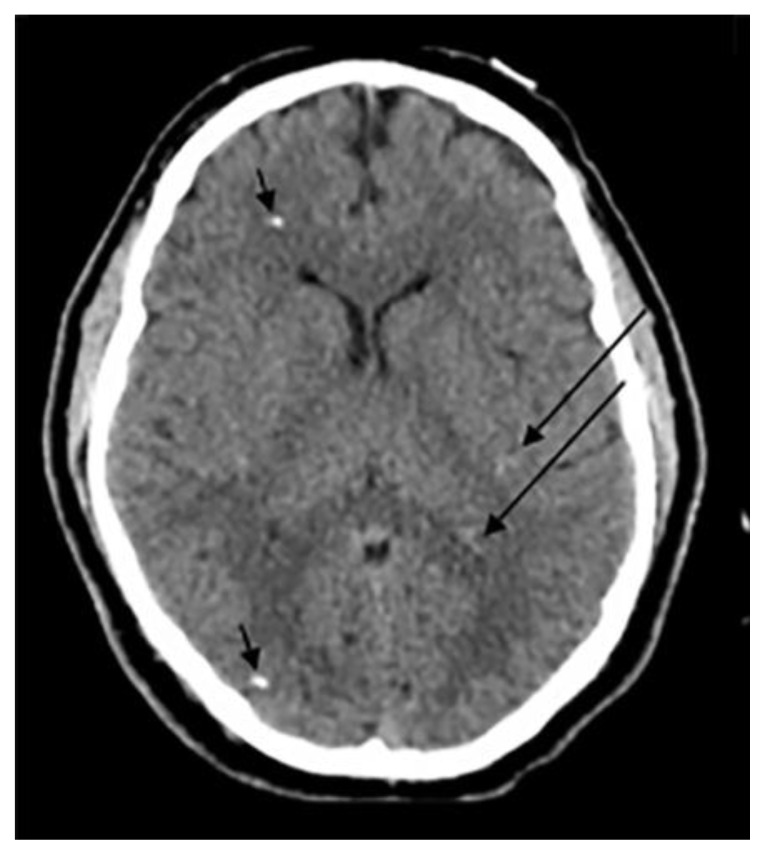
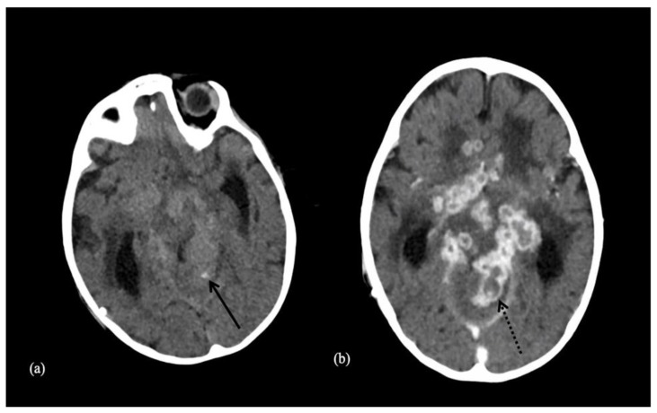
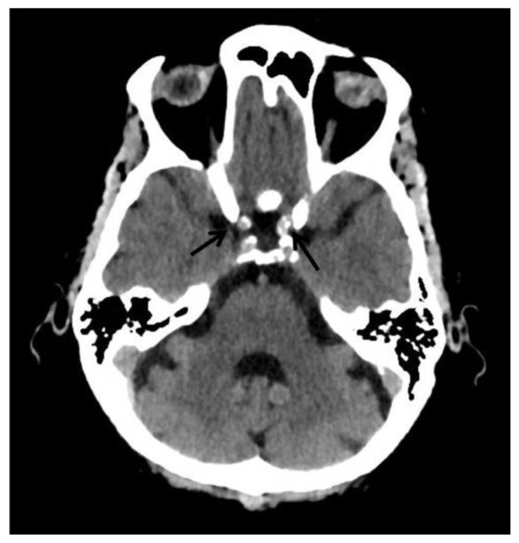
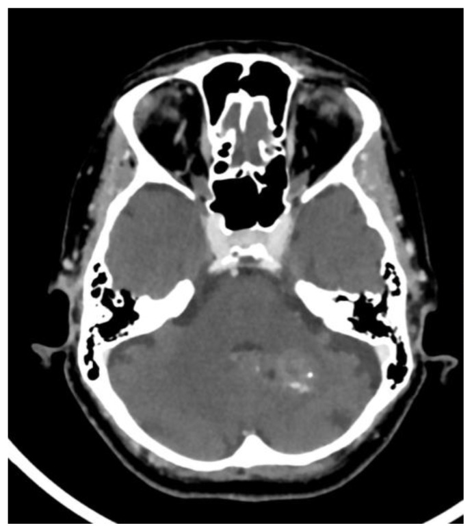
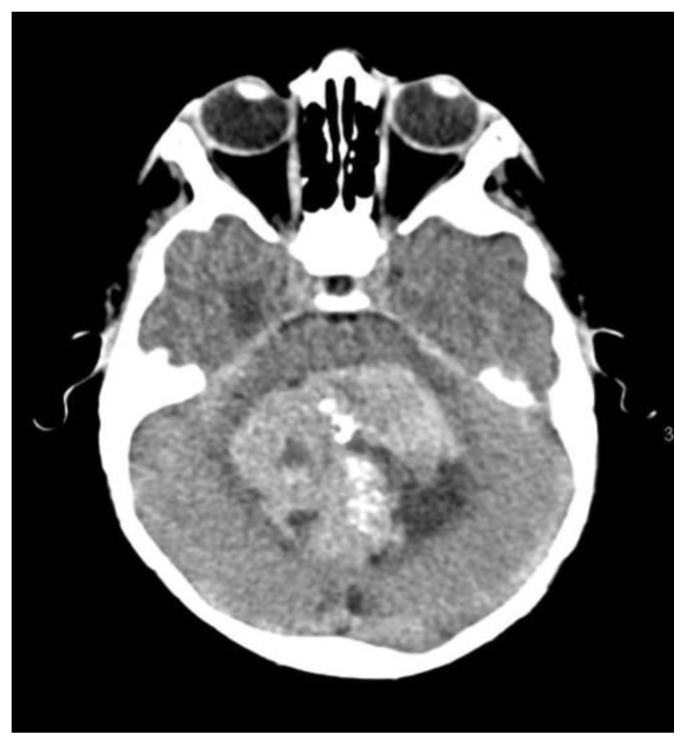
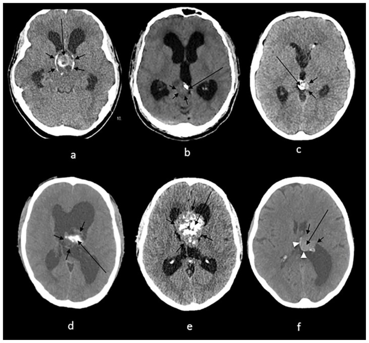
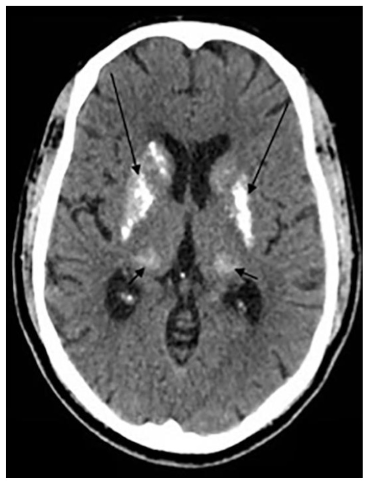
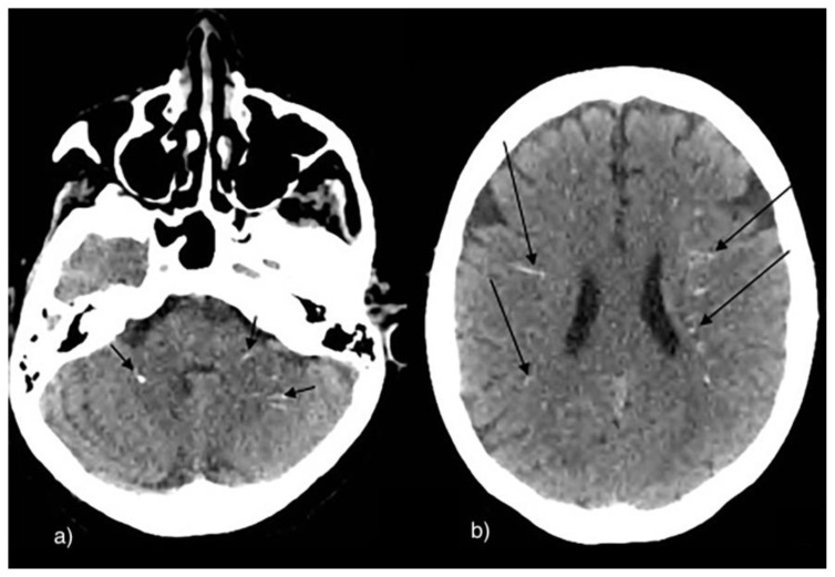
Intracranial calcifications are frequently encountered in non-contrast computed tomography scan in both adult and pediatric age groups. They refer to calcifications within the brain parenchyma or vasculature and can be classified into several major categories: physiologic/age-related, dystrophic, congenital disorders/phakomatoses, infectious, vascular, neoplastic, metabolic/endocrine, inflammatory and toxic diseases. In this updated review, we present a wide spectrum of intracranial calcifications from both pediatric and adult populations focusing on their pattern, size and location.

Keywords: Adult calcification; CT; Intracranial calcifications; Pathologic calcifications; Pediatric calcifications; Physiologic calcifications.


Figures

Figure 1
Figure 1
Examples of patterns of calcification and related terminology. (a) dots, (b) lines, (c) conglomerate or mass-like, (d) rock-like, (e) blush, (f) gyriform/band-like, (g) stippled (h) reticular.
Figure 2
Figure 2
Technique: Axial non enhanced CT, 450 mAs, 120 kV, 0.8 mm slice thickness. (a): 55-year-old female presenting to the ER post trauma. Findings: Incidental dots of calcifications in the pineal gland (arrow). (b) and (c): An 11-year-old male who presented to ER post MVA. Findings: Incidental tiny dots (less than 1 cm) of calcifications with no soft tissue component in the pineal gland (2b) and choroid plexuses (2c) (arrows), a pattern suggestive of benign/ physiologic nature. (d): 66-year-old male who presented to the ER for dizziness. Findings: Incidental dots of calcifications in the bilateral choroid plexuses (arrows). (e): 73-year-old male presenting to ER for high blood pressure and headache. Findings: Incidental dot of calcification in anterior cerebral falx (arrow). (f): 79-year-old male who presented to the ER post trauma. Findings: Incidental isolated dots of basal ganglia calcifications (arrows).
Figure 3
Figure 3
Technique: Axial non-enhanced CT, 450 mAs, 120 kV, 0.8 mm slice thickness (a): 1-month-old infant boy with Sturge-Webber syndrome. Findings: Diffuse gyral/subcortical white matter calcifications (arrows) with volume loss in the left cerebral hemisphere. (b): 6-year-old boy with tuberous sclerosis. Findings: Subependymal (short arrows) and subcoritcal tubers (long arrows), some of which are calcified. (c): 40-year-old female with NF 2. Findings: Right cerebellar hemisphere calcifications (arrow). (d): 29-year- old male with Cockayne disease. Findings: Bilateral conglomerate of calcifications in the globus pallidi, putamina, caudate nuclei and thalami (arrows) as well in the subcortical white matter (arrowheads). There is cerebral volume loss as seen by prominence of sulci and ventricles. (e): 4-year-old boy with Krabbe s disease. Findings: Blush-like calcifications within the bilateral basal ganglia (arrows) including the thalami early in the course of the disease. In addition, there is decreased bifrontal peri-ventricular density from demyelination (arrowheads). (f) 3-month-old infant girl with AGS. Findings: Conglomerate of intracranial calcifications almost equally involving the basal ganglia (arrows) including the thalami. There is slight ex-vacuo dilatation of the ventricles and slight prominence of cerebral sulci secondary to volume loss. (g): 42-year-old man with Fahr disease. Findings: Conglomerate/mass like calcifications in the bilateral basal ganglia (short arrows), bilateral cerebellar white matter and dentate nuclei (long arrows).
Figure 4
Figure 4
Technique: Axial non-enhanced CT, 450 mAs, 120 kV, 0.8 mm slice thickness. (a): 10-month-old infant boy with seizures and neonatal CMV infection. Findings: Reticular calcification pattern in the subependymal (long arrows) and periventricular region (short arrows) with mild ventriculomegaly. (b): 21-year-old female with congenital toxoplasma infection. Findings: Dots of calcification in the periventricular (long arrow) and subcortical (short arrow) regions with brain destruction, volume loss and ex-vacuo dilatation of the ventricles.
Figure 5
Figure 5
24-year- old male with previous HIV encephalitis. Technique: Axial non-enhanced CT, 450 mAs, 120 kV, 0.8 mm slice thickness. Findings: Coarse calcifications in the cerebral white matter (short arrows) and in the basal ganglia (long arrows).
Figure 6
Figure 6
33-year-old male with cysticercosis. Technique: Axial non-enhanced CT, 450 mAs, 120 kV, 0.8 mm slice thickness. Findings: Dots of calcifications in subependymal region (long arrow) and at gray to white matter interface (short arrows).
Figure 7
Figure 7
9-year-old male patient with a tuberculoma. Technique: Axial enhanced (b) and non-enhanced CT (a), 450 mAs, 120 kV, 0.8 mm slice thickness. Findings: Tuberculoma with tiny calcification on both contrast and non-contrast enhanced CT scan. Both arrows show a central calcification within the tuberculoma.
Figure 8
Figure 8
68-year-old female patient presenting with bilateral internal carotid artery atherosclerosis. Technique: Axial enhanced and non-enhanced CT, 450 mAs, 120 kV, 0.8 mm slice thickness. Findings: Bilateral internal carotid artery atherosclerosis. Arrows show atherosclerotic calcifications.
Figure 9
Figure 9
70-year-old female patient presenting with cavernous angioma. Technique: Axial enhanced and non-enhanced CT, 450 mAs, 120 kV, 0.8 mm slice thickness. Findings: Punctate calcification found in the cavernous angioma.
Figure 10
Figure 10
8-year-old male patient presenting with medulloblastoma. Technique: Axial enhanced and non-enhanced CT, 450 mAs, 120 kV, 0.8 mm slice thickness. Findings: Scattered and clumped calcifications.
Figure 11
Figure 11
Technique: Axial non enhanced CT, 450 mAs, 120 kV, 0.8 mm slice thickness. (a): 38-year-old female with craniopharyngioma. Findings: A sellar/suprasellar mass-like (long arrow) and rim calcifications (short arrows) impinging on the foramen of Monro causing hydrocephalus. (b): 20-year-old male with pineocytoma. Findings: A mass in the pineal gland (short arrows) with peripheral calcification (long arrow). (c): 13-year-old boy with pineal teratoma. Findings: Conglomerate of dense calcifications (long arrow) inside the pineal mass (short arrows). (d): 27-year-old male with intraventricular ependymoma. Findings: An irregular mass centered in the body of left lateral ventricle (short arrows) containing dense mass-like calcifications (long arrow) with secondary enlargement of the lateral ventricles. (e): 42-year-old male with central neurocytoma. Findings: A mass in the septum pellucidum (short arrows) containing conglomerates of calcifications (long arrow), obstructing the foramen of Monro and causing hydrocephalus of lateral ventricles. (f): 62-year-old female with intraventricular meningioma. Findings: Interventricular meningioma centered in posterior body of left lateral ventricle (short arrows) showing internal blush-like calcifications (long arrow) and a rim of calcifications (arrowheads) with secondary enlargement of the left occipital horn.
Figure 12
Figure 12
29-year-old male with hyperparathyroidism. Technique: Axial non enhanced CT, 450 mAs, 120 kV, 0.8 mm slice thickness. Findings: Conglomerate of calcifications in the bilateral lentiform nuclei (long arrows) and in the bilateral thalami (short arrows).
Figure 13
Figure 13
63-year-old male with hypothyroidism. Technique: Axial non-enhanced CT, 450 mAs, 120 kV, 0.8 mm slice thickness. Findings: Multiple scattered small linear calcifications in the cerebellar (short arrows) and periventricular white matter (long arrows) bilaterally.

References

    1. Livingston JH, Stivaros S, Warren D, Crow YJ. Intracranial calcification in childhood: a review of aetiologies and recognizable phenotypes. Developmental Medicine & Child Neurology. 2014 Jul;56(7):612–26. - PubMed
    1. Deng H, Zheng W, Jankovic J. Genetics and molecular biology of brain calcification. Ageing research reviews. 2015 Jul 1;22:20–38. - PubMed
    1. Yalcin A, Ceylan M, Bayraktutan OF, Sonkaya AR, Yuce I. Age and gender related prevalence of intracranial calcifications in CT imaging; data from 12,000 healthy subjects. Journal of chemical neuroanatomy. 2016 Dec 1;78:20–4. - PubMed
    1. Grech R, Grech S, Mizzi A. Intracranial Calcifications: A Pictorial Review. The neuroradiology journal. 2012 Aug;25(4):427–51. - PubMed
    1. Whitehead MT, Oh C, Raju A, Choudhri AF. Physiologic pineal region, choroid plexus, and dural calcifications in the first decade of life. American Journal of Neuroradiology. 2015 Mar 1;36(3):575–80. - PMC - PubMed