Review
. 2019 Aug;9(Suppl 1):S116-S130.
doi: 10.21037/cdt.2018.11.02.

Vascular imaging in renal donors

Affiliations
Review

Vascular imaging in renal donors

Ayaz Aghayev et al. Cardiovasc Diagn Ther. 2019 Aug.

Abstract

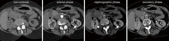
Imaging plays a crucial role in pre-transplant evaluation to enhance the probability of a successful outcome. Its aim is to define kidney and vascular anatomy and to assess potential pathologies. Each modality has advantages and disadvantages. Computed tomography angiography (CTA) is the most commonly used imaging modality, however, magnetic resonance angiography (MRA) can be used in selected cases. The purpose of this review article is to provide an overview of available imaging modalities, their benefits, risks, advantages, and disadvantages. Imaging findings that indicate particular anomalies and pathologies that may affect living renal donor selection will be discussed.

Keywords: Renal donor; transplantation; vascular imaging.


Conflict of interest statement

Conflicts of Interest: The authors have no conflicts of interest to declare.

Figures

Figure 1
Figure 1
Four-phase CT images demonstrate a tiny stone in the right kidney upper pole (arrow), which is not appreciated on arterial, nephrographic or excretory phases.
Figure 2
Figure 2
Four-phase CT images show an 8 mm cyst in the left kidney (arrow), which is best appreciated in the nephrographic phase. On arterial phase only, the image it can be masked by the nonenhanced medulla. A tiny cyst (arrowhead) in the right kidney is also well-seen on nephrographic phase.
Figure 3
Figure 3
The left (A) and the right (B) kidney measurements on multiplanar reformatted (MPR) CT images.
Figure 4
Figure 4
The left (A) and the right (B) renal artery pre-hilar arterial branching measurements on curved multiplanar reformatted (cMPR) CT images. The arrows in the left lower corner demonstrate the distance to the branching.
Figure 5
Figure 5
Maximum intensity projection (MIP) CTA image through the kidney shows main (arrows) and small accessory (arrowhead) as well as aberrant renal (curved arrow) arteries.
Figure 6
Figure 6
Curved multiplanar reformatted (cMPR) CTA images of the venous system demonstrate distance to the left (A) and right (B) renal vein confluences as well as gonadal (C) and adrenal veins (D) drainages into the left renal vein. Arrows indicates gonadal vein in (C), adrenal gland verin in (D).
Figure 7
Figure 7
Volume-rendered 3D CT (A) and MRI (B) excretory phase images demonstrate the normal anatomy of the collecting system.
Figure 8
Figure 8
Volume-rendered 3D CTA image of the abdominal aorta and renal arteries (A) shows early branching (arrow) of the left renal artery, and curved multiplanar reformatted (cMPR) image of the same artery (B) demonstrates the distance (arrows) from the aorta to the level of the branching. Similarly, volume-rendered 3D MRA image of the abdominal aorta and right renal artery (C) demonstrate early branching (black and white arrows), and CTA and MRA images are comparable.
Figure 9
Figure 9
Small accessory left renal artery (arrow) is seen in the volume-rendered 3D CTA image (A). Similarly, in a volume-rendered 3D (B) and maximum intensity projection (MIP) (C) MRA images demonstrate bilateral accessory renal arteries.
Figure 10
Figure 10
Volume-rendered 3D CTA image shows two aberrant (polar) renal arteries supplying the left kidney one is arising from the aorta (arrow), and the second one is originating from the left common iliac artery (arrowhead).
Figure 11
Figure 11
A patient excluded from the renal donor list due to three right renal arteries, and two left renal arteries.
Figure 12
Figure 12
Maximum intensity projection (MIP) (A) and volume-rendered 3D (B) MRA images demonstrate severe stenosis (arrows) at the proximal segment of the right renal artery due to atherosclerotic disease.
Figure 13
Figure 13
Volume-rendered 3D (A) and maximum intensity projection (MIP) (B) CTA images demonstrate incidentally detected fibromuscular dysplasia (FMD) both renal arteries (arrows). Similarly, a patient with incidentally detected FMD on MIP MRA image (C) and was confirmed on conventional angiography image (D). Arrows in (C) indicates FMD in the right renal artery.
Figure 14
Figure 14
Maximum intensity projection (MIP) CTA image (A) demonstrates a circumaortic left renal vein; the branch anterior to the abdominal aorta (arrow) is draining into the proximal portion of the inferior vena cava (IVC), whereas the branch posterior to the abdominal aorta (arrowhead) is draining to the distal portion of the IVC. MIP CTA image demonstrates retroaortic left renal vein (arrow) (B).
Figure 15
Figure 15
Maximum intensity projection (MIP) MRA image (A) demonstrate duplicated IVC and the left renal vein (arrow) is draining into the left-sided IVC (arrowhead). Coronal MIP MRA image (B) demonstrates rare venous anomaly of the azygous continuation of IVC, and left renal vein is draining into the azygous at the level of the heart.
Figure 16
Figure 16
Volume-rendered 3D CT image (A) shows a slightly ptotic right kidney. Volume-rendered 3D CT image (B) demonstrates partial duplex collecting system on the left (arrows), and fuses at the level of the midabdomen.
Figure 17
Figure 17
Axial CT image through the abdomen (A) demonstrates the congenital absence of the left kidney with a unicornuate uterus (B), and a CT image through the abdomen (C) in another patient shows a horseshoe kidney.

References

    1. Tullius SG, Rabb H. Improving the Supply and Quality of Deceased-Donor Organs for Transplantation. N Engl J Med 2018;378:1920-9. 10.1056/NEJMra1507080 - DOI - PubMed
    1. Simforoosh N, Basiri A, Tabibi A, et al. Comparison of laparoscopic and open donor nephrectomy: a randomized controlled trial. BJU International 2005;95:851-5. 10.1111/j.1464-410X.2005.05415.x - DOI - PubMed
    1. Sebastià C, Peri L, Salvador R, et al. Multidetector CT of living renal donors: lessons learned from surgeons. Radiographics 2010;30:1875-90. 10.1148/rg.307105032 - DOI - PubMed
    1. Davis CL, Delmonico FL. Living-donor kidney transplantation: a review of the current practices for the live donor. J Am Soc Nephrol 2005;16:2098-110. 10.1681/ASN.2004100824 - DOI - PubMed
    1. Smith D, Chudgar A, Daly B, et al. Evaluation of potential renal transplant recipients with computed tomography angiography. Arch Surg 2012;147:1114-22. 10.1001/archsurg.2012.1466 - DOI - PubMed