Review
. 2019 Aug;9(Suppl 1):S174-S182.
doi: 10.21037/cdt.2019.07.08.

Lower extremity arteries

Affiliations
Review

Lower extremity arteries

Manish Kumar Yadav et al. Cardiovasc Diagn Ther. 2019 Aug.

Abstract

Lower extremity arteries play vital role of supplying blood to the extremity bone, muscles, tendons and nerves to maintain the mobility of the body. These arteries may get involved with a number of disease processes which restrict the optimal functioning of the limb. The knowledge of various diseases, clinical presentation, appearance on various imaging modalities and segments of involvement helps one to clinch the diagnosis. It is of paramount importance for imaging clinician to apply the correct imaging tool based on the clinical question which is facilitated by know how of the advantages and limitation of each of these imaging modalities. This article focuses on lower extremity arteries, its anatomy, various imaging modalities and common disease conditions affecting the lower limb arteries.

Keywords: Lower extremity arteries; computerized tomography; digital subtraction angiography; doppler; magnetic resonance imaging; ultrasound.


Conflict of interest statement

Conflicts of Interest: The authors have no conflicts of interest to declare.

Figures

Figure 1
Figure 1
Digital subtraction angiogram at aortic bifurcation. (A) Digital Subtraction angiogram at the level of aortic bifurcation (black arrow) showing the division of aorta into right and left common iliac arteries; (B) same DSA with magnification demonstrating the median sacral artery (blue arrow).
Figure 2
Figure 2
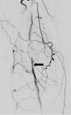
Digital subtraction angiogram of a 64 years old patient with severe calf claudication. Angiogram shows rich collateral network around the knee joint reforming the P2 segment of the popliteal artery (black arrow).
Figure 3
Figure 3
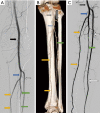
Angiogram demonstrating the anatomy of popliteal and tibial arteries. (A) Digital subtraction angiogram of left leg showing a normal popliteal artery (black arrow) and its division into anterior tibial (white arrows) and tibioperoneal trunk (blue arrow). The tibioperoneal trunk further divides into peroneal artery (green arrows) and posterior tibial artery (yellow arrows); (B) CT angiogram volume rendered technique posterior view demonstrating the popliteal artery and its division. Note the site of anterior tibial artery piercing the interosseous membrane to move anteriorly (grey arrow); (C) DSA of below knee tibial vessels.
Figure 4
Figure 4
Atherosclerotic disease on imaging. (A) CT plain Sagittal reformat of hip and thigh region of a 65 years old lady with poorly controlled diabetic and chronic renal failure patient showing severe vessel wall calcifications in common femoral, superficial femoral and profunda femoris artery (white arrows); (B) DSA of another 71 years old patient with diabetes and hypertension showing diffuse atherosclerotic changes in the form of multiple eccentric plaques (black arrows); (C) below knee angiogram of same patient as (B) showing single vessel runoff in the form of anterior tibial artery, peroneal artery is occluded just beyond its origin and posterior tibial artery shows diffuse disease.
Figure 5
Figure 5
CT angiogram of 65 years old patient who presented with acute pain in right leg. (A) Popliteal artery aneurysm (black arrow) as seen on axial CT image and (B) in sagittal CT MIP image. Note the occlusion of the artery just beyond the popliteal aneurysm.
Figure 6
Figure 6
Thirty-four years old female with right lower extremity claudication. (A) MRA coronal in dorsiflexion position showing severe compression of popliteal artery from gastrocnemius muscle; (B) axial fs spgr showing bulky gastrocnemius muscle (white arrow).
Figure 7
Figure 7
Fifty-nine years old male with claudication, Cystic adventitial disease of popliteal artery (A) CT VRT showing occlusion of the right popliteal artery (white arrow), absence of atherosclerotic disease in rest of the vasculature; (B) coronal MIP of the CT angiogram showing well defined cystic lesion causing extrinsic severe compression (yellow arrow); (C) sagittal MIP of CT angiogram showing non enhancing cystic lesion causing extrinsic compression (yellow arrow).
Figure 8
Figure 8
CT angiogram volume rendered technique of a 31 years old male with diffuse angiomatosis of left lower limb with multiple arteriovenous malformations in the limb. (A) Dilated left lower limb arteries and veins (white arrow), note the filling of the left lower limb veins whereas the right lower limb veins are not opacified yet. The size of the left lower limb arteries is grossly enlarged compared to normal sized contralateral vessels. Multiple sites of arteriovenous malformation and arteriovenous shuntings (yellow arrow) (B) note the large venous sac in the draining vein (blue arrow); (C) grossly hypertrophied left lower limb compared to the normal right lower limb.
Figure 9
Figure 9
Imaging (CT) in vasculitis. (A) CT axial venous phase shows occlusion of infrarenal aorta with wall thickening and enhancement of the wall (black arrow); (B) CT angiogram VRT showing the infrarenal aortic occlusion (white arrows) with distal reformation of bilateral external iliac artery (blue arrows).
Figure 10
Figure 10
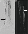
Thromboangitis obliterans. (A) Digital Subtraction angiogram of a 27 years old patient, smoker with right great toe ulcer and rest pain. Occlusion of the anterior tibial artery with cork screw collaterals (black arrow); (B) occlusion of posterior tibial artery with cork screw collaterals (white arrow).

Similar articles

Cited by

References

    1. Butler P, Mitchell AWM, Ellis H, Healy JC. Applied radiological anatomy. Cambridge University Press; 2012:400.
    1. Bajzer Christopher T. Arterial supply of the lower extremities - Guide to Peripheral and Cerebrovascular Intervention - NCBI Bookshelf. Remedica Pub, 2004:291.
    1. Scheinert D, Werner M, Scheinert S, et al. Treatment of Complex Atherosclerotic Popliteal Artery Disease With a New Self-Expanding Interwoven Nitinol Stent: 12-Month Results of the Leipzig SUPERA Popliteal Artery Stent Registry. JACC Cardiovasc Interv 2013;6:65-71. 10.1016/j.jcin.2012.09.011 - DOI - PubMed
    1. Koshy CG, Chacko BR, Nidugala Keshava S, et al. Diagnostic accuracy of color Doppler imaging in the evaluation of peripheral arterial disease as compared To digital subtraction angiography. Vasc Dis Manag 2009;6:2-9.
    1. Kohler TR, Nance DR, Cramer MM, et al. Duplex scanning for diagnosis of aortoiliac and femoropopliteal disease: a prospective study. Circulation 1987;76:1074-80. 10.1161/01.CIR.76.5.1074 - DOI - PubMed