Review
. 2019 Sep;8(Suppl 3):S168-S177.
doi: 10.21037/gs.2019.06.01.

Neuroblastoma image-defined risk factors in adrenal neuroblastoma: role of radiologist

Affiliations
Review

Neuroblastoma image-defined risk factors in adrenal neuroblastoma: role of radiologist

Cecilia Lanza et al. Gland Surg. 2019 Sep.

Abstract

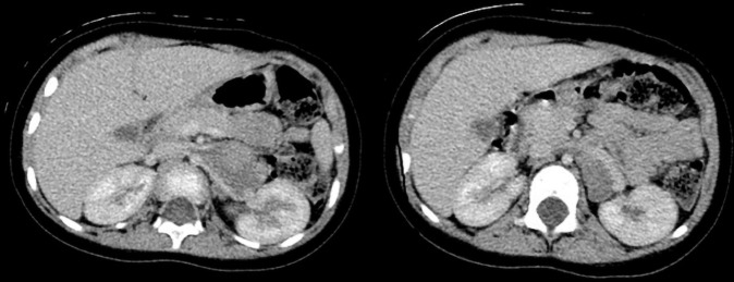
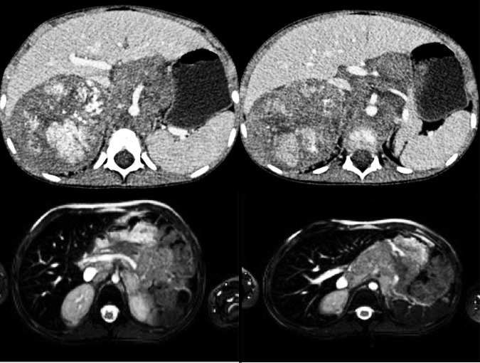
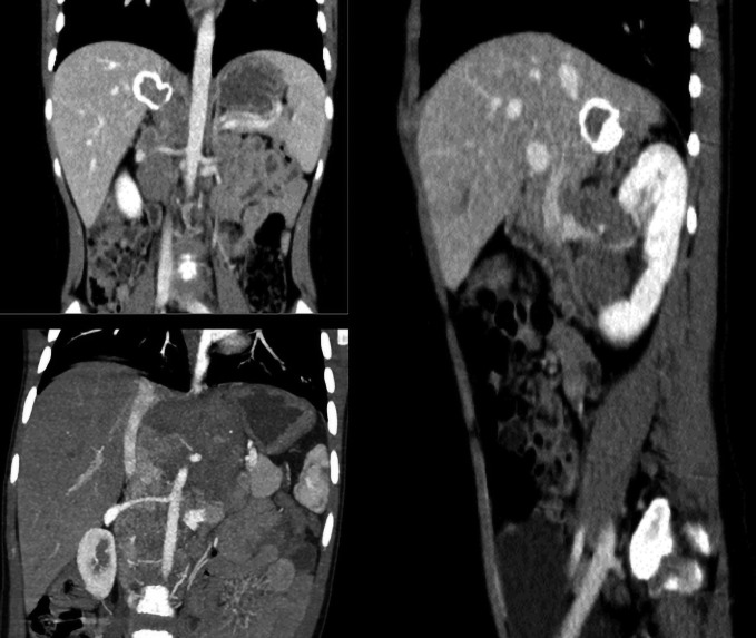
Neuroblastoma, one of the most common extracranial solid malignancies in children, is often localized in the adrenal glands (49%). The staging system for prognostic purpose was one of the first points of disagreement, which led to the International Neuroblastoma Staging System (INSS) of 1986, revised in 1989, which relies on surgical staging. The limit of this classification was the different surgical resection, also done at interval times from diagnosis. To overcome this difficulty, a new staging system was made based on preoperative imaging by the International Neuroblastoma Risk Group (INRG) in 2009. This new staging system uses 20 Image-Defined Risk Factors (IDRFs) across multiple organ systems. The scope of this IDRFs is to predict surgical outcomes and, in addition with clinical data, to provide risk stratification. The INRG Staging System (INRGSS) relies on Imaging-Defined Risk Factors (IDRFs) that are determined before surgery or other therapy. With the application of the INRGSS the radiologist's role in staging children with neuroblastoma increased. The review provides an overview of the INRGSS and the IDRFs in adrenal neuroblastoma.

Keywords: Adrenal gland; INRG Staging System (INRGSS); Imaging-Defined Risk Factors (IDRFs); International Neuroblastoma Risk Group (INRG); neuroblastoma (NBL).


Conflict of interest statement

Conflicts of Interest: The authors have no conflicts of interest to declare.

Figures

Figure 1
Figure 1
Typical findings in adrenal neuroblastoma. (A) Ultrasound shows heterogeneous solid mass which cystic component; (B) computed tomography after contrast medium administration shows large heterogeneous lesion with calcifications arising in right adrenal gland; (C) magnetic resonance T2W fat sat sequence shows hyperintense retroperitoneal lesion involving aorta and goes beyond the median line; (D) evidence of multiple liver metastatic lesions (Stage MS) on computed tomography (I) and ultrasound (II) studies.
Figure 2
Figure 2
CT after contrast medium administration. Contact: there’s involvement of less than 50% of the vessel’s circumference of aorta, splenic artery and celiac axis. CT, computed tomography.
Figure 3
Figure 3
CT after contrast medium administration. Contact with the renal artery, vein and renal pelvis. CT, computed tomography.
Figure 4
Figure 4
CT and MR. Encasement: there isn’t a visible layer of normal tissue between tumor and celiac trunk and portal vein. There’s an involvement of more than 50% of the circumference of the celiac artery. CT, computed tomography; MR, magnetic resonance.
Figure 5
Figure 5
CT after contrast medium administration. Infiltration of vena cava: malignant tissue grows into vena cava with loss defined margins. CT, computed tomography.
Figure 6
Figure 6
CT after contrast medium administration. Invasion with encasement of the renal vessels. CT, computed tomography.
Figure 7
Figure 7
MR and CT: encasement of the celiac axis, superior mesenteric arteries, aorta, and renal pedicles. MR, magnetic resonance; CT, computed tomography.

References

    1. Dumba M, Jaward N, McHung K. Neuroblastoma and nephroblastoma: a radiological review. Cancer Imaging 2015;15:5. 10.1186/s40644-015-0040-6 - DOI - PMC - PubMed
    1. Liu W, Zheng J, Li Q. Application of imaging modalities for evaluating neuroblastoma. J Pediatr Endocrinol Metab 2013;26:1015-20. 10.1515/jpem-2013-0117 - DOI - PubMed
    1. Brisse HJ, McCarville MB, Granata C, et al. International Neuroblastoma Risk Group Project. Guidelines for imaging and staging of neuroblastic tumors: consensus report from the International Neuroblastoma Risk Group Project. Radiology 2011;261:243-57. 10.1148/radiol.11101352 - DOI - PubMed
    1. Müller-Schulte E, Kurlemann G, Harder A. Tobacco, alcohol and illicit drugs during pregnancy and risk of neuroblastoma: systematic review. Arch Dis Child Fetal Neonatal Ed 2018;103:F467-73. 10.1136/archdischild-2017-313615 - DOI - PubMed
    1. Fletcher JI, Ziegler DS, Trahair TN, et al. Too many targets, not enough patients: rethinking neuroblastoma clinical trials; Nat Rev Cancer 2018;18:389-400. 10.1038/s41568-018-0003-x - DOI - PubMed

LinkOut - more resources