Review
. 2019 Sep;8(Suppl 3):S178-S187.
doi: 10.21037/gs.2019.04.06.

Groove pancreatitis: a challenging imaging diagnosis

Affiliations
Review

Groove pancreatitis: a challenging imaging diagnosis

Gloria Addeo et al. Gland Surg. 2019 Sep.

Abstract

Groove pancreatitis (GP) is an uncommon form of chronic pancreatitis (CP) involving the space between duodenum, pancreatic head and common bile duct (CBD) known as pancreatic-duodenal groove. Although an association with long-standing ethanol assumption is reported a definite etiology of GP is unknown. Since thickening of the duodenal wall, pancreatic head enlargement, CBD stricture and dilatation of pancreatic duct system are common findings the differential diagnosis with pancreatic head neoplasm by means of imaging can be challenging. However, some imaging findings such as fibrotic changes of the pancreatic groove and presence of duodenal wall cysts may suggest the correct diagnosis. In this paper we review clinical and imaging features of GP with emphasis on computed tomography (CT) and magnetic resonance imaging (MRI) findings.

Keywords: Groove pancreatitis (GP); computed tomography (CT); magnetic resonance cholangiopancreatography (MRCP); magnetic resonance imaging (MRI).


Conflict of interest statement

Conflicts of Interest: The authors have no conflicts of interest to declare.

Figures

Figure 1
Figure 1
This picture shows the pancreatic-duodenal groove (asterisks), a “theoretic space” between the medial duodenal wall and the head of the pancreas.
Figure 2
Figure 2
Axial contrast-enhanced CT image in a 54-year-old male affected by pure-form of groove pancreatitis showing the pancreatic-duodenal artery (PDA) (arrowhead) that represent an anatomic landmark ox axial image to distinguish the duodenal groove respect to the pancreatic head. Duodenal groove is lateral respect the PDA. In this case the duodenal groove in widened due to the presence of hypodense curvilinear tissue (asterisk). Medial duodenal wall is also thickened with intramural cyst (arrow). Dilatation of MPD is also visible. MPD, main pancreatic duct; CT, computed tomography.
Figure 3
Figure 3
Groove pancreatitis, segmental form: contrast-enhanced CT axial image (A) and coronal reconstruction (B) showing “sheet-like” hypodense and “patchy” enhanced soft tissue in the pancreatic-duodenal groove (asterisks) involving the pancreatic head with tapering of common bile duct (arrowhead). CT, computed tomography.
Figure 4
Figure 4
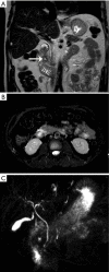
Early stage of pure groove pancreatitis at MRI: coronal T2-weighted image (A), axial T2-weighted acquired with fat-saturation technique (B) and MRCP (C) showing intramural cyst of the medial duodenal wall (arrow) and widening of the duodenal pancreatic groove due to the presence of “sheet-like” soft tissue with high signal intensity on T2-weighted-fat-saturated image (asterisk) due to soft-tissue edema. MRCP image confirm the widening of the pancreatic-duodenal-groove (asterisk) and depict the lack of common bile duce and pancreatic duct dilatation. MRI, magnetic resonance imaging; MRCP, magnetic resonance cholangiopancreatography.
Figure 5
Figure 5
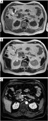
Groove pancreatitis at MRI: axial-unenhanced T1-weighted fat-saturated image (A), axial T2-weighted image (B) and delayed-enhanced-T1-weighted-fat-satured image (C). Images show medial duodenal wall thickening with low signal intensity on T1 weighted image (arrow); cystic intramural changes of the medial duodenal wall better visible on T2-weighted image (arrow) and delayed enhancement of fibrotic tissue of the duodenal wall (arrow). MRI, magnetic resonance imaging.
Figure 6
Figure 6
Groove pancreatitis pure form. Axial T1-weighted image (A), axial T2-weighted image (B) and contrast-enhanced axial T1-weighted fat-saturated image (C) showing soft tissue mass in the pancreatic-duodenal groove (arrows) with low signal intensity on T1-weighted image, low signal intensity respect to pancreatic gland on T2-weightd image and “patchy” enhancement. Arrowhead indicate the pancreatic-duodenal-artery.

References

    1. Becker V. Bauchspeicheldrüse. In: Doerr W, Seifert G, Uhlinger E. editors. Spezielle pathologische Anatomie. Vol 4. Berlin: Springer, 1973:252-445.
    1. Stolte M, Weiss W, Volkholz H, et al. A special form of segmental pancreatitis: groove pancreatitis. Hepatogastroenterology 1982;29:198-208. - PubMed
    1. Becker V, Mischke U. Groove pancreatitis. Int J Pancreatol 1991;10:173-82. - PubMed
    1. Adsay NV, Zamboni G. Paraduodenal pancreatitis: a clinicopathologically distinct entity unifying 'cystic dystrophy of heterotopic pancreas', 'paraduodenal wall cyst', and 'groove pancreatitis'. Semin Diagn Pathol 2004;21:247-54. 10.1053/j.semdp.2005.07.005 - DOI - PubMed
    1. Blasbalg R, Baroni RH, Costa DN, et al. MRI features of groove pancreatitis. AJR Am J Roentgenol 2007;189:73-80. 10.2214/AJR.06.1244 - DOI - PubMed