

High-resolution crystal structures of a myxobacterial phytochrome at cryo and room temperatures
- PMID: 31559319
- PMCID: PMC6748860
- DOI: 10.1063/1.5120527
High-resolution crystal structures of a myxobacterial phytochrome at cryo and room temperatures
Abstract
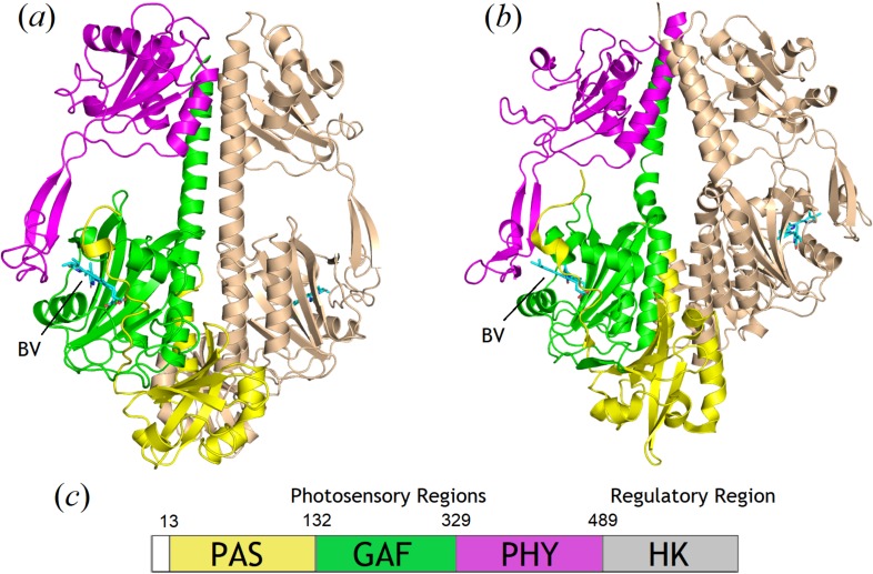
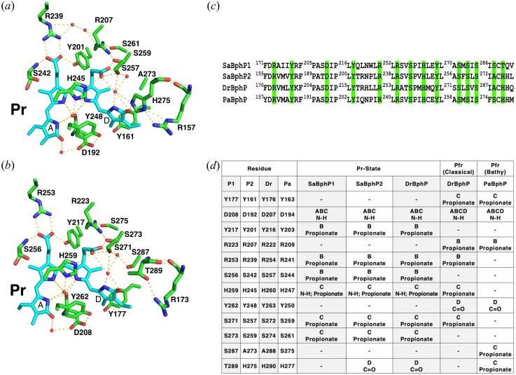
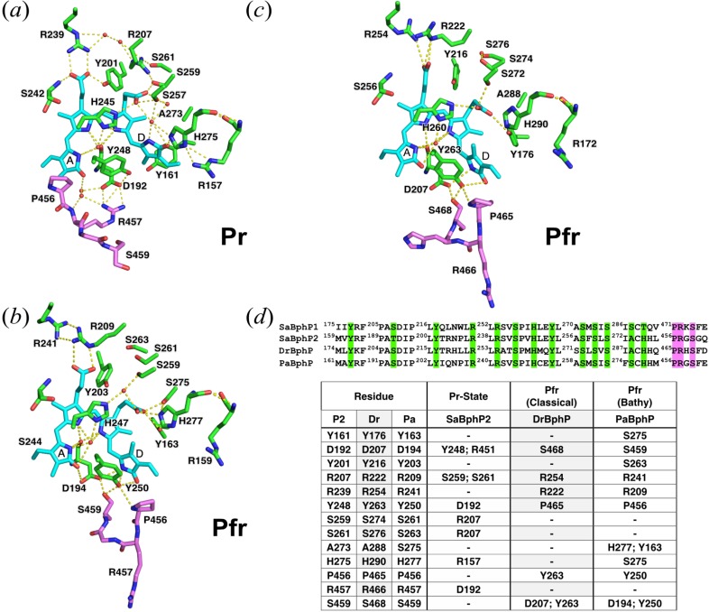
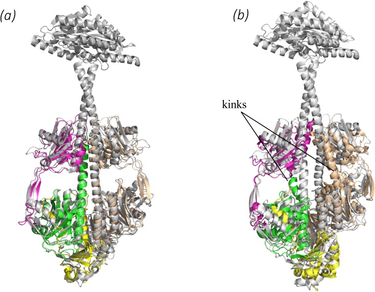
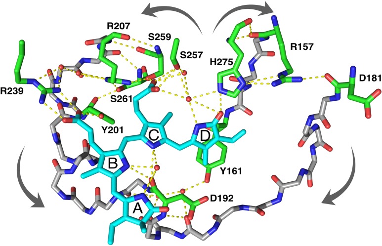
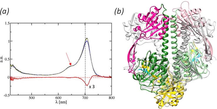
Phytochromes (PHYs) are photoreceptor proteins first discovered in plants, where they control a variety of photomorphogenesis events. PHYs as photochromic proteins can reversibly switch between two distinct states: a red light (Pr) and a far-red light (Pfr) absorbing form. The discovery of Bacteriophytochromes (BphPs) in nonphotosynthetic bacteria has opened new frontiers in our understanding of the mechanisms by which these natural photoswitches can control single cell development, although the role of BphPs in vivo remains largely unknown. BphPs are dimeric proteins that consist of a photosensory core module (PCM) and an enzymatic domain, often a histidine kinase. The PCM is composed of three domains (PAS, GAF, and PHY). It holds a covalently bound open-chain tetrapyrrole (biliverdin, BV) chromophore. Upon absorption of light, the double bond between BV rings C and D isomerizes and reversibly switches the protein between Pr and Pfr states. We report crystal structures of the wild-type and mutant (His275Thr) forms of the canonical BphP from the nonphotosynthetic myxobacterium Stigmatella aurantiaca (SaBphP2) in the Pr state. Structures were determined at 1.65 Å and 2.2 Å (respectively), the highest resolution of any PCM construct to date. We also report the room temperature wild-type structure of the same protein determined at 2.1 Å at the SPring-8 Angstrom Compact free electron LAser (SACLA), Japan. Our results not only highlight and confirm important amino acids near the chromophore that play a role in Pr-Pfr photoconversion but also describe the signal transduction into the PHY domain which moves across tens of angstroms after the light stimulus.
Figures
Similar articles
-
Structural basis for light control of cell development revealed by crystal structures of a myxobacterial phytochrome.IUCrJ. 2018 Aug 29;5(Pt 5):619-634. doi: 10.1107/S2052252518010631. eCollection 2018 Sep 1. IUCrJ. 2018. PMID: 30224965 Free PMC article.
-
Dynamics and efficiency of photoswitching in biliverdin-binding phytochromes.Photochem Photobiol Sci. 2019 Oct 9;18(10):2484-2496. doi: 10.1039/c9pp00264b. Photochem Photobiol Sci. 2019. PMID: 31418445
-
Signal Transduction in an Enzymatic Photoreceptor Revealed by Cryo-Electron Microscopy.bioRxiv [Preprint]. 2023 Nov 9:2023.11.08.566274. doi: 10.1101/2023.11.08.566274. bioRxiv. 2023. PMID: 37986774 Free PMC article. Preprint.
-
Phytochrome three-dimensional structures and functions.Biochem Soc Trans. 2010 Apr;38(2):710-6. doi: 10.1042/BST0380710. Biochem Soc Trans. 2010. PMID: 20298248 Review.
-
The system of phytochromes: photobiophysics and photobiochemistry in vivo.Membr Cell Biol. 1998;12(5):691-720. Membr Cell Biol. 1998. PMID: 10379648 Review.
Cited by
-
Blue and red in the protein world: Photoactive yellow protein and phytochromes as revealed by time-resolved crystallography.Struct Dyn. 2024 Jan 31;11(1):014701. doi: 10.1063/4.0000233. eCollection 2024 Jan. Struct Dyn. 2024. PMID: 38304445 Free PMC article.
-
Trends in coordination of rhenium organometallic complexes in the Protein Data Bank.IUCrJ. 2022 Feb 25;9(Pt 2):180-193. doi: 10.1107/S2052252522000665. eCollection 2022 Mar 1. IUCrJ. 2022. PMID: 35371500 Free PMC article. Review.
-
On the Role of the Conserved Histidine at the Chromophore Isomerization Site in Phytochromes.J Phys Chem B. 2021 Dec 23;125(50):13696-13709. doi: 10.1021/acs.jpcb.1c08245. Epub 2021 Nov 29. J Phys Chem B. 2021. PMID: 34843240 Free PMC article.
-
Structures of myxobacterial phytochrome revealed by cryo-EM using the Spotiton technique and with x-ray crystallography.Struct Dyn. 2025 May 1;12(3):034701. doi: 10.1063/4.0000301. eCollection 2025 May. Struct Dyn. 2025. PMID: 40322674 Free PMC article.
-
Phylogenetic Analysis with Prediction of Cofactor or Ligand Binding for Pseudomonas aeruginosa PAS and Cache Domains.Microbiol Spectr. 2021 Dec 22;9(3):e0102621. doi: 10.1128/spectrum.01026-21. Epub 2021 Dec 22. Microbiol Spectr. 2021. PMID: 34937179 Free PMC article.
References
-
- Woitowich N. C., Halavaty A. S., Waltz P., Kupitz C., Valera J., Tracy G. et al., “ Structural basis for light control of cell development revealed by crystal structures of a myxobacterial phytochrome,” Int. Union Crystallogr. J. 5(Pt. 5), 619–634 (2018).10.1107/S2052252518010631 - DOI - PMC - PubMed
Grants and funding
LinkOut - more resources
Full Text Sources
Research Materials