

Recursive hierarchical embedding in vision is impaired by posterior middle temporal gyrus lesions
- PMID: 31560064
- PMCID: PMC6763734
- DOI: 10.1093/brain/awz242
Recursive hierarchical embedding in vision is impaired by posterior middle temporal gyrus lesions
Abstract
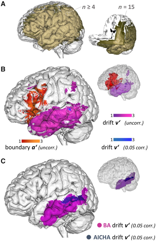
The generation of hierarchical structures is central to language, music and complex action. Understanding this capacity and its potential impairments requires mapping its underlying cognitive processes to the respective neuronal underpinnings. In language, left inferior frontal gyrus and left posterior temporal cortex (superior temporal sulcus/middle temporal gyrus) are considered hubs for syntactic processing. However, it is unclear whether these regions support computations specific to language or more generally support analyses of hierarchical structure. Here, we address this issue by investigating hierarchical processing in a non-linguistic task. We test the ability to represent recursive hierarchical embedding in the visual domain by contrasting a recursion task with an iteration task. The recursion task requires participants to correctly identify continuations of a hierarchy generating procedure, while the iteration task applies a serial procedure that does not generate new hierarchical levels. In a lesion-based approach, we asked 44 patients with left hemispheric chronic brain lesion to perform recursion and iteration tasks. We modelled accuracies and response times with a drift diffusion model and for each participant obtained parametric estimates for the velocity of information accumulation (drift rates) and for the amount of information accumulated before a decision (boundary separation). We then used these estimates in lesion-behaviour analyses to investigate how brain lesions affect specific aspects of recursive hierarchical embedding. We found that lesions in the posterior temporal cortex decreased drift rate in recursive hierarchical embedding, suggesting an impaired process of rule extraction from recursive structures. Moreover, lesions in inferior temporal gyrus decreased boundary separation. The latter finding does not survive conservative correction but suggests a shift in the decision criterion. As patients also participated in a grammar comprehension experiment, we performed explorative correlation-analyses and found that visual and linguistic recursive hierarchical embedding accuracies are correlated when the latter is instantiated as sentences with two nested embedding levels. While the roles of the inferior temporal gyrus and posterior temporal cortex in linguistic processes are well established, here we show that posterior temporal cortex lesions slow information accumulation (drift rate) in the visual domain. This suggests that posterior temporal cortex is essential to acquire the (knowledge) representations necessary to parse recursive hierarchical embedding in visual structures, a finding mimicking language acquisition in young children. On the contrary, inferior frontal gyrus lesions seem to affect recursive hierarchical embedding processing by interfering with more general cognitive control (boundary separation). This interesting separation of roles, rooted on a domain-general taxonomy, raises the question of whether such cognitive framing is also applicable to other domains.
Keywords: hierarchy; inferior frontal gyrus; lesion; syntax; visuospatial.
© The Author(s) (2019). Published by Oxford University Press on behalf of the Guarantors of Brain.
Figures


Similar articles
-
Fractal image perception provides novel insights into hierarchical cognition.Neuroimage. 2014 Aug 1;96:300-8. doi: 10.1016/j.neuroimage.2014.03.064. Epub 2014 Mar 31. Neuroimage. 2014. PMID: 24699014
-
A novel approach to investigate recursion and iteration in visual hierarchical processing.Behav Res Methods. 2016 Dec;48(4):1421-1442. doi: 10.3758/s13428-015-0657-1. Behav Res Methods. 2016. PMID: 26487047
-
Recursion in action: An fMRI study on the generation of new hierarchical levels in motor sequences.Hum Brain Mapp. 2019 Jun 15;40(9):2623-2638. doi: 10.1002/hbm.24549. Epub 2019 Mar 5. Hum Brain Mapp. 2019. PMID: 30834624 Free PMC article.
-
A rostro-caudal gradient of structured sequence processing in the left inferior frontal gyrus.Philos Trans R Soc Lond B Biol Sci. 2012 Jul 19;367(1598):2023-32. doi: 10.1098/rstb.2012.0009. Philos Trans R Soc Lond B Biol Sci. 2012. PMID: 22688637 Free PMC article. Review.
-
Hierarchy processing in human neurobiology: how specific is it?Philos Trans R Soc Lond B Biol Sci. 2020 Jan 6;375(1789):20180391. doi: 10.1098/rstb.2018.0391. Epub 2019 Nov 18. Philos Trans R Soc Lond B Biol Sci. 2020. PMID: 31735144 Free PMC article. Review.
Cited by
-
Hierarchical Structure in Sequence Processing: How to Measure It and Determine Its Neural Implementation.Top Cogn Sci. 2020 Jul;12(3):910-924. doi: 10.1111/tops.12442. Epub 2019 Jul 30. Top Cogn Sci. 2020. PMID: 31364310 Free PMC article. Review.
-
Intraoperative cortical localization of music and language reveals signatures of structural complexity in posterior temporal cortex.iScience. 2023 Jun 28;26(7):107223. doi: 10.1016/j.isci.2023.107223. eCollection 2023 Jul 21. iScience. 2023. PMID: 37485361 Free PMC article.
-
Alterations of the alpha rhythm in visual snow syndrome: a case-control study.J Headache Pain. 2024 Apr 8;25(1):53. doi: 10.1186/s10194-024-01754-x. J Headache Pain. 2024. PMID: 38584260 Free PMC article.
-
Gray matter asymmetry in asymptomatic carotid stenosis.Hum Brain Mapp. 2021 Dec 1;42(17):5665-5676. doi: 10.1002/hbm.25645. Epub 2021 Sep 9. Hum Brain Mapp. 2021. PMID: 34498785 Free PMC article.
-
Finding Hierarchical Structure in Binary Sequences: Evidence from Lindenmayer Grammar Learning.Cogn Sci. 2023 Jan;47(1):e13242. doi: 10.1111/cogs.13242. Cogn Sci. 2023. PMID: 36655988 Free PMC article.
References
-
- Ashburner J, Friston KJ. Unified segmentation. Neuroimage 2005; 26: 839–51. - PubMed
-
- Bates D, Martin M, Bolker B, Walker S. lme4: Linear Mixed-Effects Models Using Eigen and S4. R Package Version 1.0–6 [Internet]. 2014. http://cran.r-project.org/package=lme4.
-
- Bates E, Wilson SM, Saygin AP, Dick F, Sereno MI, Knight RT et al. . Voxel-based lesion-symptom mapping. Nat Neurosci 2003; 6: 448. - PubMed
-
- Berwick RC, Chomsky N. Why only us: language and evolution. Cambridge: The MIT Press; 2015.
-
- Berwick RC, Friederici AD, Chomsky N, Bolhuis JJ. Evolution, brain, and the nature of language. Trends Cogn Sci 2013; 17: 98. - PubMed
MeSH terms
LinkOut - more resources
Full Text Sources
Research Materials
