Review
. 2019 Nov-Dec;170(8):321-337.
doi: 10.1016/j.resmic.2019.09.004. Epub 2019 Sep 24.

An integrated transport mechanism of the maltose ABC importer

Affiliations
Review

An integrated transport mechanism of the maltose ABC importer

Rebecca Mächtel et al. Res Microbiol. 2019 Nov-Dec.

Abstract

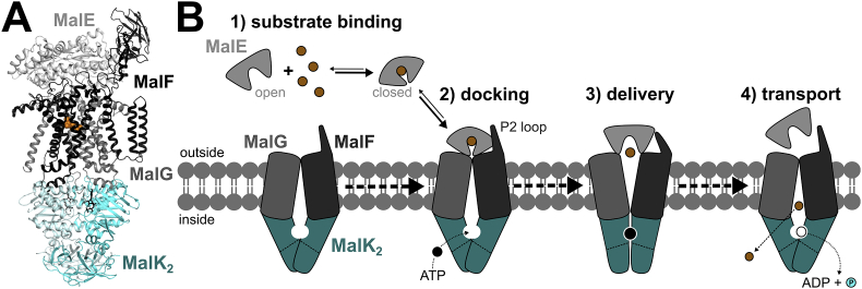
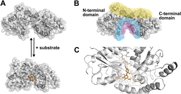
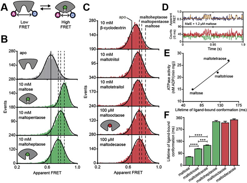
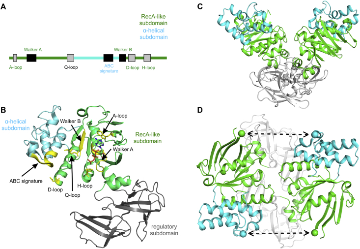
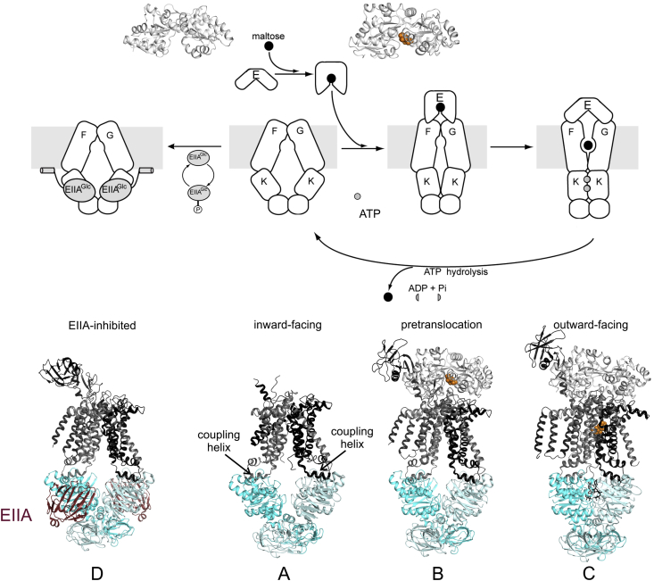
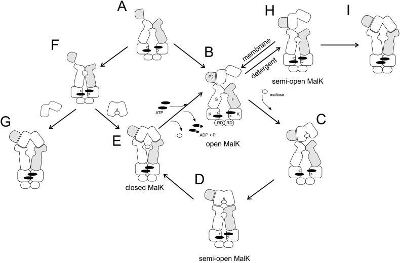
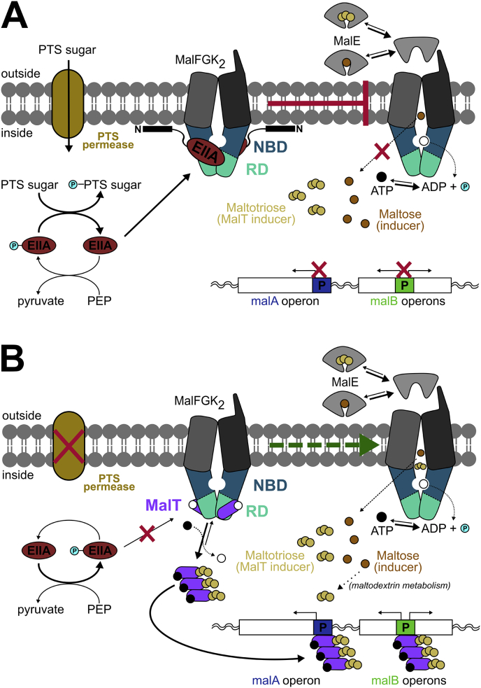
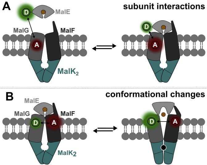
ATP-binding cassette (ABC) transporters use the energy of ATP hydrolysis to transport a large diversity of molecules actively across biological membranes. A combination of biochemical, biophysical, and structural studies has established the maltose transporter MalFGK2 as one of the best characterized proteins of the ABC family. MalF and MalG are the transmembrane domains, and two MalKs form a homodimer of nucleotide-binding domains. A periplasmic maltose-binding protein (MalE) delivers maltose and other maltodextrins to the transporter, and triggers its ATPase activity. Substrate import occurs in a unidirectional manner by ATP-driven conformational changes in MalK2 that allow alternating access of the substrate-binding site in MalF to each side of the membrane. In this review, we present an integrated molecular mechanism of the transport process considering all currently available information. Furthermore, we summarize remaining inconsistencies and outline possible future routes to decipher the full mechanistic details of transport by MalEFGK2 complex and that of related importer systems.

Keywords: ABC transporter; EPR spectroscopy; Importer; Single molecule fluorescence; Substrate-binding protein; smFRET.


Figures

Fig. 1
Fig. 1
Maltose transport by MalEFGK2. A, Crystal structure of MalEFGK2 in complex with ATP and maltose. MalE is docked onto MalFG in the outward ATP-occluded state, and a maltose molecule is colored in orange spheres (PDB: 2R6G); B, Schematic of the import mechanism of maltose. The four steps depicted are discussed in detail in this review. (For interpretation of the references to color in this figure legend, the reader is referred to the Web version of this article.)
Fig. 2
Fig. 2
MalE structure and conformational states.A, Crystal structures of MalE, without substrate bound (PDB: 1OMP) and with maltose bound (PDB: 1ANF). The “closure” around the ligand within the binding pocket led to the term “venus flytrap mechanism”. B, Regions often mentioned in the literature are indicated in different colors: hinge region or balancing interface (yellow), binding pocket (red) and lip region (blue). As there is no consensus about the concrete residues assembling each region, the cartoon was kept simple and is just an indication including residues named in different publications. C, MalE binding pocket with maltose (shown in orange) bound. Red dashed lines indicate hydrogen bonds to subsite B (D14, K15, E111) or both, subsite B and C (E153, Y155). Subsite A and D are not involved and do not have relevance for maltose binding in E. coli MalE (PDB: 1ANF). (For interpretation of the references to color in this figure legend, the reader is referred to the Web version of this article.)
Fig. 3
Fig. 3
smFRET to study the closure of MalE in the presence of various sugars. A, smFRET assay for characterization of MalE closure by using fluorophore proximity for B, Transported (green) and C, non-transported (red) ligands. D, Real-time monitoring of ligand-driven conformational changes in isolated MalE by smFRET with example of maltose. E, Positive correlation between ligand release times derived from data similar to that of D from isolated MalE in comparison to ATPase activity of MalEFGK2. F, Ligand-release times of various transported (maltose, maltotriose, maltotetraose, maltoheptaose) and non-transported ligands (maltooctaose, maltodecaose). All panels are reprinted from de Boer et al. . (For interpretation of the references to color in this figure legend, the reader is referred to the Web version of this article.)
Fig. 4
Fig. 4
MalK structure with conserved motifs. A, Conserved motifs in the NBD of MalK. B, ATP-bound MalK structure (PDB: 1Q12) showing the subdomains and conserved motifs (in yellow) . C, Open MalK dimer viewed from the side (PDB: 1Q1E). D, Open MalK dimer viewed from the top. The green and cyan spheres represent the V16 and R129 residues, respectively, that were used to monitor distance changes during closure of the dimer interface via EPR spectroscopy . Closure of the MalK dimer is indicated by the dashed lines. (For interpretation of the references to color in this figure legend, the reader is referred to the Web version of this article.)
Fig. 5
Fig. 5
Structures of the maltose transporter in various states of the transport cycle. Crystal structures of MalE are shown in its open (maltose free; PDB: 1OMP) and closed (maltose bound; PDB: 1ANF) conformations. The maltose transporter was crystallized in several conformational states: Inward-facing MalFGK2 in its resting state (PDB: 3FH6, see A), MalEFGK2 in a pretranslocation state (PDB: 3PV0 or 3PUZ, see B), MalEFGK2 outward-facing state (PDB: 2R6G, see C) and MalFGK2 in inward-facing state inhibited by its EIIAGlc regulatory protein (PDB: 4JBW, see D). A scheme of maltose delivery by MalE to MalFGK2 is displayed based on the crystal structures.
Fig. 6
Fig. 6
Integrated mechanism of maltose transport based on structural and biochemical studies. Cartoons A, D, E are directly derived from crystal structures (A, PDB: 3FH6; D, 3PUZ; E, 3RLF or 2R6G). EPR studies from Davidson and Bordignon’s groups suggested that the main conformational route occurs through ABCDEB, or ACDEB if maltose is highly available , , , , . Step C is probably transient but is depicted for clarity. In detergent, the ATPase activity of MalEFGK2 was insensitive to the presence of maltose, and EPR studies suggested that this was due to the increased ability of MalEFGK2 in detergent to transition through H in the absence of maltose . Note that conformations I and G are also observed by EPR and cross-linking studies . A different model was proposed by the group of Duong, in which ATP alone induces the outward-facing conformation . However, this model was later refined to a route through F, in which MalK2 is asymmetric and semi-open in the presence of ATP . The interaction of liganded MalE then stabilizes this conformation (similar as in D) to promote the conversion toward E, a route that is consistent with EPR data, especially if F is a small or transient population.
Fig. 7
Fig. 7
Mechanism for the control of maltose uptake. A, Schematic model of the inducer exclusion mechanism. PTS permeases facilitate PTS sugar uptake into the cell. EIIAglc is dephosphorylated, interacts with NBD and RD from different monomers of MalK, and is anchored to the inner leaflet of the membrane. The maltose transporter MalFGK2 is thus trapped in an inactive conformation and maltodextrins uptake is inhibited. Consequently, maltodextrins (e.g. maltose, maltotriose) cannot further induce the maltose system by promoting the expression of malA and malB genes. B, Representation of the MalT positive regulation. In the absence of PTS sugars, the equilibrium of EIIAglc is shifted towards its phosphorylated state (EIIAglc-P) which cannot interact with MalK; therefore, transport can occur. Under these conditions, maltose induces the expression of malA and malB regions through its channeling into a metabolic pathway mediated by MalT-inducible enzymes. MalT is released from MalK and can be activated by (cognate) maltotriose imported or produced from the maltodextrin metabolism. MalT multimerization (upon its activation) allows its binding to the malA and malB promoters thereby inducing gene transcription. Of note, the divergent operon that belongs to the malA region and contains the malT gene is not shown, since it does not belong to the maltose regulon. Moreover, MalT-inducible operons located in other loci than malA and malB are also not shown for simplicity.
Fig. 8
Fig. 8
Proposed future smFRET studies of MalEFGK2. A, Visualization of one of the many interactions that occur between subunits (complex formation) using smFRET. Depicted assay would monitor interactions between MalE and MalF or MalG to understand further the docking process. B, Study of intra- or interdomain conformational changes via FRET. Figure adapted from Mächtel et al. .
Fig. 9
Fig. 9
Proposed future single transporter recordings using periplasmic substrate binding protein-based fluorescent biosensors. A, Schematic of the proposed experimental set-up for single-transporter recordings. Single-transporters are reconstituted into proteoliposomes at low protein-to-lipid ratios to have one (or none) transporter per liposome, providing positive and negative control experiments. The position of the liposome can be detected via a fluorescent marker. Liposomes are filled with a large quantity of fluorescent sensors such as SBPs that detect changes in the concentration of the substrate, here via increase in their signal. B, Assay design for use of MalE as maltose-sensor using an environmentally-sensitive fluorophore (blue: IANBD). Fluorescence spectra at increasing maltose concentration from 0 (low fluorescence) to 1 mM maltose (high fluorescence). C, Scheme of the TIRF-microscope with laser box and dual-view allowing simultaneous recording of two emission channels, exemplified by data from a lipid marker DiD and luminal dsDNA-fluorescein. D, Example data of fluorescent markers in lumen and the liposome leaflet as schematics for expected signals upon single-transporter recordings. (For interpretation of the references to color in this figure legend, the reader is referred to the Web version of this article.)

References

    1. Davidson A.L., Dassa E., Orelle C., Chen J. Structure, function, and evolution of bacterial ATP-binding cassette systems. Microbiol Mol Biol Rev. 2008;72:317–364. [table of contents] - PMC - PubMed
    1. Dassa E. Natural history of ABC systems: not only transporters. Essays Biochem. 2011;50:19–42. - PubMed
    1. Gouridis G., Hetzert B., Kiosze-Becker K., de Boer M., Heinemann H., Nurenberg-Goloub E. ABCE1 controls ribosome recycling by an asymmetric dynamic conformational equilibrium. Cell Rep. 2019;28:723–734 e6. - PMC - PubMed
    1. Locher K.P. Mechanistic diversity in ATP-binding cassette (ABC) transporters. Nat Struct Mol Biol. 2016;23:487–493. - PubMed
    1. ter Beek J., Guskov A., Slotboom D.J. Structural diversity of ABC transporters. J Gen Physiol. 2014;143:419–435. - PMC - PubMed

MeSH terms

LinkOut - more resources