

Simultaneous quantification of estrogens and glucocorticoids in human adipose tissue by liquid-chromatography-tandem mass spectrometry
- PMID: 31561001
- PMCID: PMC7099401
- DOI: 10.1016/j.jsbmb.2019.105476
Simultaneous quantification of estrogens and glucocorticoids in human adipose tissue by liquid-chromatography-tandem mass spectrometry
Abstract
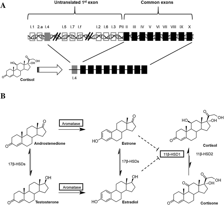
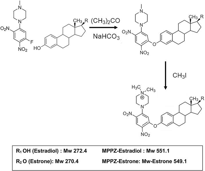
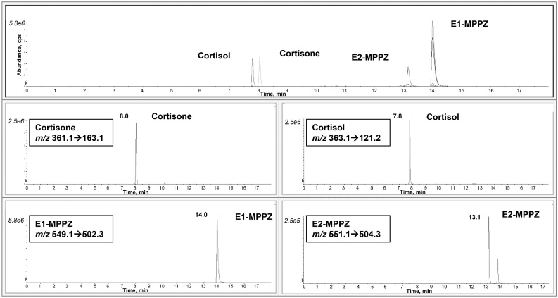
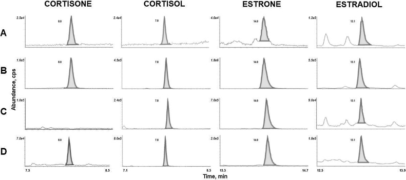
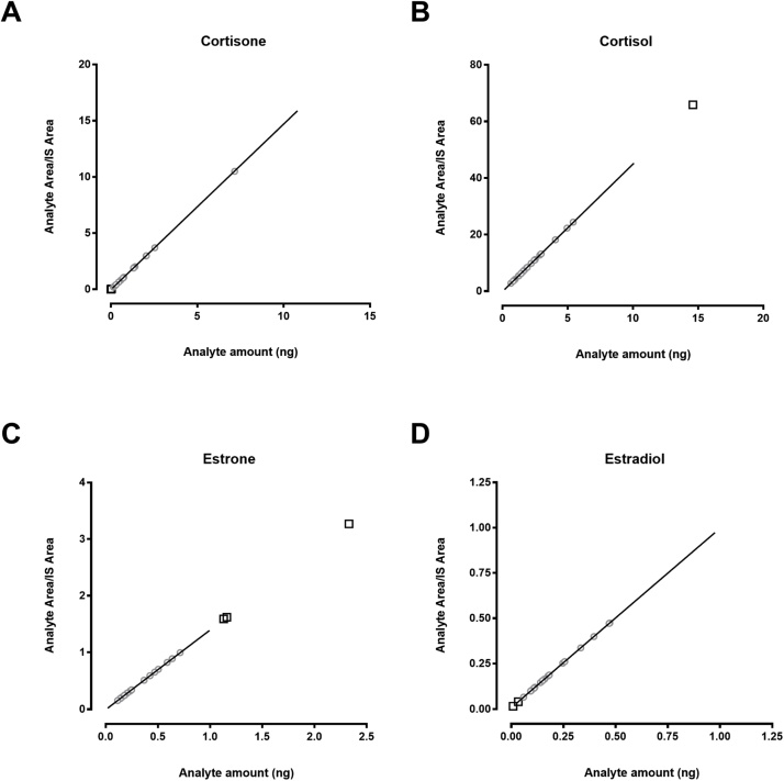
The presence of estrogens, androgens and glucocorticoids as well as their receptors and steroid converting enzymes in adipose tissue has been established. Their contribution to diseases such as obesity, diabetes and hormone-dependent cancers is an active area of research. Our objective was to develop a LC-MS/MS method to quantify bioactive estrogens and glucocorticoids simultaneously in human adipose tissue. Estrogens and glucocorticoids were extracted from adipose tissue samples using solid-phase extraction. Estrogens were derivatized using 1-(2,4-dinitro-5-fluorophenyl)-4-methylpiperazine (PPZ) and methyl iodide to generate a permanently charged molecule (MPPZ). Steroids were separated and quantified by LC-MS/MS. The limit of quantitation for the steroids was between 15 and 100 pg per sample. Accuracy and precision were acceptable (<20%). Using this method, estradiol, estrone, cortisone and cortisol were quantified in adipose tissue from women with and without breast cancer. This novel assay of estrogens and glucocorticoids by LC-MS/MS coupled with derivatization allowed simultaneous quantification of a panel of steroids in human adipose tissue across the endogenous range of concentrations encountered in health and disease.
Keywords: Adipose; Cortisol; Cortisone; Derivatization; Estradiol; Estrone.
Copyright © 2019 The Authors. Published by Elsevier Ltd.. All rights reserved.
Conflict of interest statement
AT is the recipient of research grant support from Johnson & Johnson Medical Companies and Medtronic for studies unrelated to this publication.
Figures
References
-
- Tchernof A., Mansour M.F., Pelletier M., Boulet M.M., Nadeau M., Luu-The V. Updated survey of the steroid-converting enzymes in human adipose tissues. J. Steroid Biochem. Mol. Biol. 2015;147:56–69. - PubMed
-
- Yamatani H., Takahashi K., Yoshida T., Takata K., Kurachi H. Association of estrogen with glucocorticoid levels in visceral fat in postmenopausal women. Menopause. 2013;20(4):437–442. - PubMed
-
- Zhao Y., Nichols J.E., Bulun S.E., Mendelson C.R., Simpson E.R. Aromatase P450 gene expression in human adipose tissue. Role of a Jak/STAT pathway in regulation of the adipose-specific promoter. J. Biol. Chem. 1995;270(27):16449–16457. - PubMed
-
- Jamieson P.M., Nyirenda M.J., Walker B.R., Chapman K.E., Seckl J.R. Interactions between oestradiol and glucocorticoid regulatory effects on liver-specific glucocorticoid-inducible genes: possible evidence for a role of hepatic 11beta-hydroxysteroid dehydrogenase type 1. J. Endocrinol. 1999;160(1):103–109. - PubMed
