. 2019 Sep 26;7(10):397.
doi: 10.3390/microorganisms7100397.

Myxozoan Adhesion and Virulence: Ceratonova shasta on the Move

Affiliations

Myxozoan Adhesion and Virulence: Ceratonova shasta on the Move

Gema Alama-Bermejo et al. Microorganisms. .

Abstract

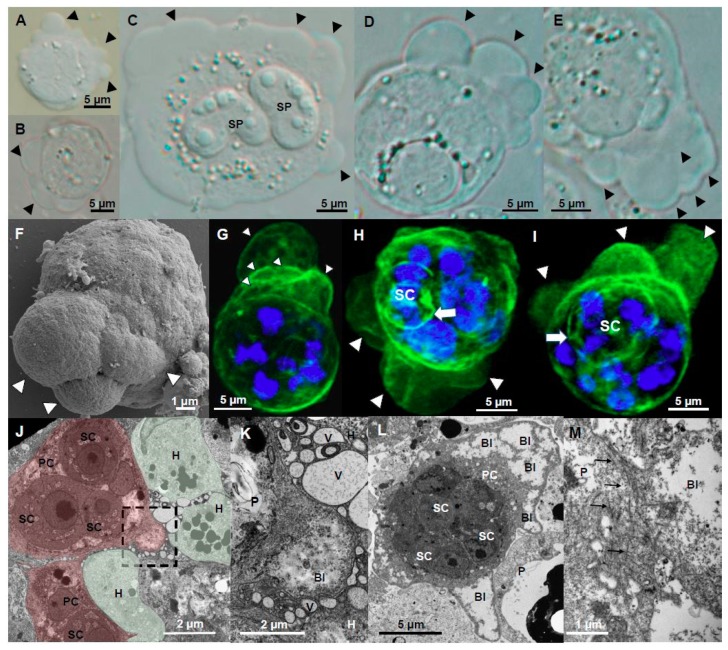
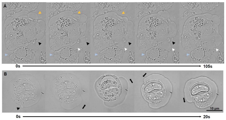
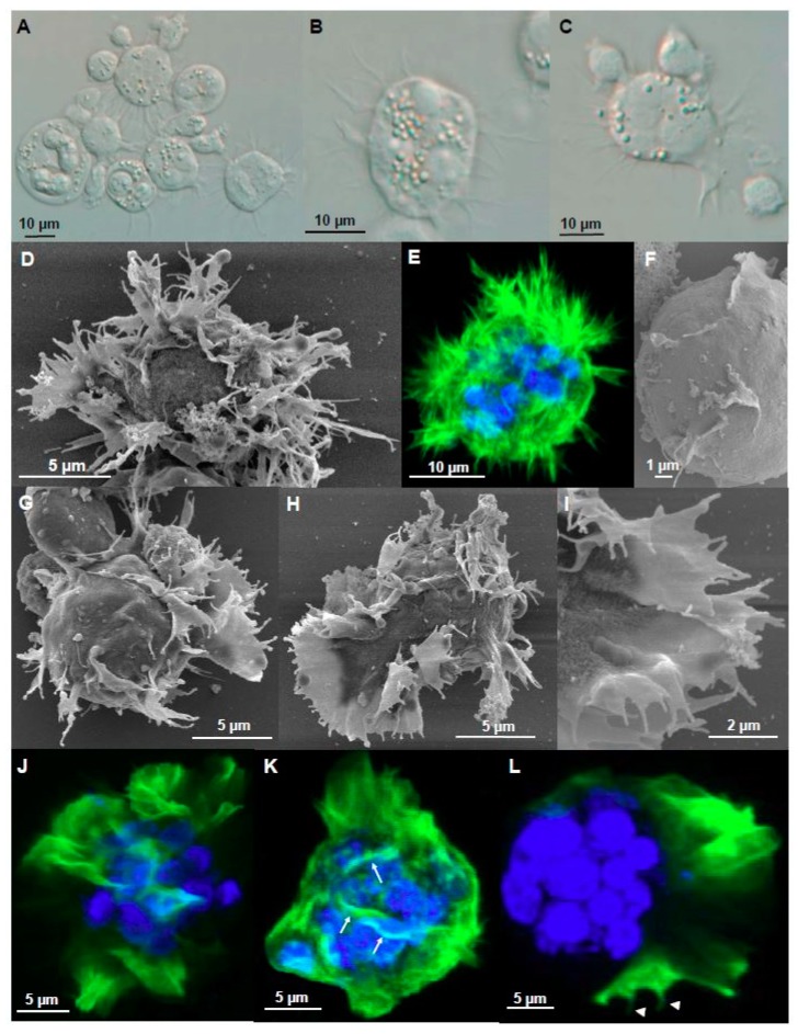
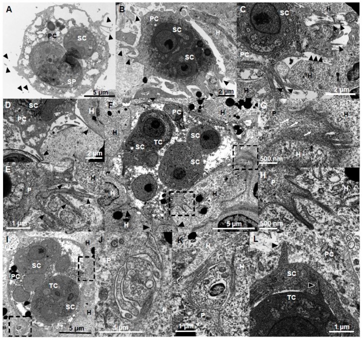
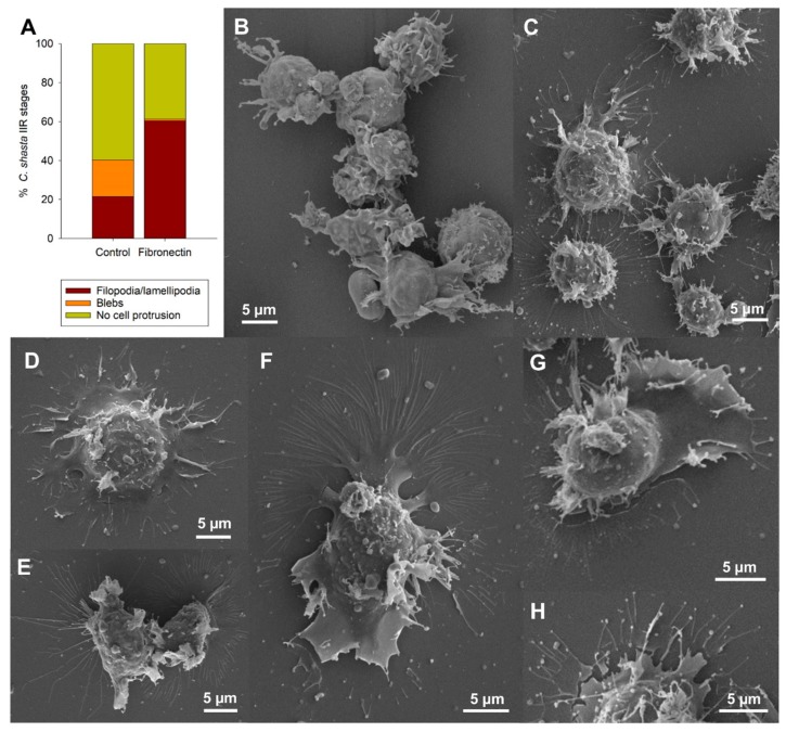
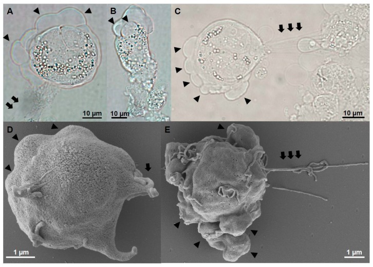
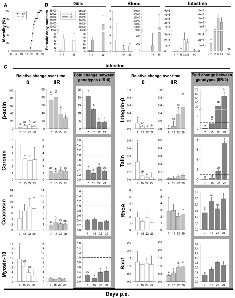
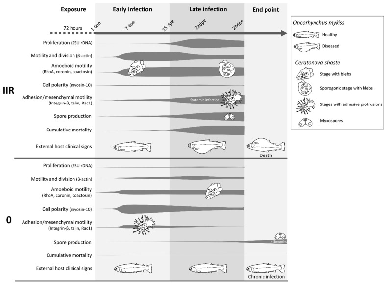
Motility factors are fundamental for parasite invasion, migration, proliferation and immune evasion and thus can influence parasitic disease pathogenesis and virulence. Salmonid enteronecrosis is caused by a myxozoan (Phylum Cnidarian) parasite, Ceratonova shasta. Three parasite genotypes (0, I, II) occur, with varying degrees of virulence in its host, making it a good model for examining the role of motility in virulence. We compare C. shasta cell motility between genotypes and describe how the cellular protrusions interact with the host. We support these observations with motility gene expression analyses. C. shasta stages can move by single or combined used of filopodia, lamellipodia and blebs, with different behaviors such as static adhesion, crawling or blebbing, some previously unobserved in myxozoans. C. shasta stages showed high flexibility of switching between different morphotypes, suggesting a high capacity to adapt to their microenvironment. Exposure to fibronectin showed that C. shasta stages have extraordinary adhesive affinities to glycoprotein components of the extracellular matrix (ECM). When comparing C. shasta genotypes 0 (low virulence, no mortality) and IIR (high virulence, high mortality) infections in rainbow trout, major differences were observed with regard to their migration to the target organ, gene expression patterns and proliferation rate in the host. IIR is characterized by rapid multiplication and fast amoeboid bleb-based migration to the gut, where adhesion (mediated by integrin-β and talin), ECM disruption and virulent systemic dispersion of the parasite causes massive pathology. Genotype 0 is characterized by low proliferation rates, slow directional and early adhesive migration and localized, non-destructive development in the gut. We conclude that parasite adhesion drives virulence in C. shasta and that effectors, such as integrins, reveal themselves as attractive therapeutic targets in a group of parasites for which no effective treatments are known.

Keywords: blebbing; cell protrusion; integrin beta; motility factors; myxozoan adhesion; rainbow trout.


Conflict of interest statement

The authors declare no conflict of interest.

Figures

Figure 1
Figure 1
Bleb protrusions on Ceratonova shasta genotype IIR stages in rainbow trout. (A,B) Small presporogonic stages showing active blebbing. (CE) Sporogonic stages showing active polarized blebbing, in (C) stage contains two myxospores. (D,E) Overlapping blebs, concentrated at one side of the stage. (F) External appearance of 3 overlapping blebs. (GI) F-actin distribution on blebbing stages. (G) Overlapping blebs. (H,I) Blebs of different sizes, probably showing blebs at different stages of their life cycle. Notice secondary cells, probably containing tertiary cells, that showed F-actin labelled membranes. (J) Parasite stage (in red) containing three secondary cells visible in the caeca surrounded by host cells (in green) and other parasite stages, showing a hemispherical bleb (square) being protruded between host cells. Notice the lack of cytoplasmic content in the bleb. (K) Detail of the bleb on J, notice the bleb is surrounded by host vesicles. (L) Parasite stage containing three secondary cells in the intestine showing profuse blebbing; notice the lack of cytoplasmic content of the blebs. (M) Base of a bleb in a liver parasite stage, with abundant actin filaments. Head arrows: blebs, White arrow, F-actin in secondary cell. Black arrows, actin filaments. A, F, Bile; BE, GI, Ascites; J,K Caeca; L, Intestine; M, liver. AE Light microscopy, F SEM, GI CLSM (Green-F-actin, phalloidin; blue-nucleic acids, DAPI), JM TEM. SP-spore; P-parasite; H-host; PC-primary cell; SC-secondary cell; Bl-bleb; V-vesicle.
Figure 2
Figure 2
Light microscopy time-lapse series of blebbing stages of Ceratonova shasta genotype IIR from ascites of rainbow trout. (A) Polarized blebbing (total time elapsed 105 s, images every 21 s), head arrows point to surface positions of the stages where blebs are expanding and rectracting, with physical overlap of several blebs simultaneously. (B) Non-polarized blebbing or circus movement (total time elapsed 20 s, images every 5 s): a bleb expansion initiated and propagated along the circumference of the sporogonic stage (with 2 mature spores), fully circumnavigating the parasite.
Figure 3
Figure 3
Filopodia and lamellipodia of adhesive stages of Ceratonova shasta genotype IIR in rainbow trout. (A) Presporogonic and sporogonic developmental stages showing abundant static filopodia. (B) Radially distributed filopodia on a stage. (C) Sheet-like lamellipodia with filopodia on the lamellipodia border and also all over the body of the stage. (D) 3D radiating pattern of filopodia. (E) F-actin distribution on a 3D radiating pattern of filopodia. (F) Detail of the parasite surface with small crests. (G,H) Parasite stages with sheet-like lamellipodia and small filopodia projecting in the external margin. Notice the sheet-like lamellipodia located on one side, with opposing single and ramified filopodia over the rest of the parasite body. (I) Detail external margin of lamellipodia with small filopodia on the external border. (JL) F-actin distribution on stages with lamellipodia. (K) Stage showing F-actin rich small crests (arrows) on the surface. (L) Small F-actin rich filopodia (head arrows) on the external margin of the lamellipodia. All stages were collected from ascites. AC Light microscopy, D, FI SEM, E, JL CLSM (Green-F-actin, phalloidin; blue-nucleic acids, DAPI).
Figure 4
Figure 4
Ultrastructure (TEM) of cell protrusions of adhesive stages of Ceratonova shasta genotype IIR in rainbow trout. (A) Sporogonic stage, containing a forming spore and a secondary cell, showing small thin filopodia protruding from the primary cell. (B) Parasite stage containing two secondary cells with abundant pseudopodia being projected into the ECM. (C) Plasmodia showing a large sheet-like lamellipodia and filopodia protruding into a degraded ECM. (D) Parasite stage protruding two long and thin filopodia between host cells. (E) Group of filopodia being projected from a parasite. (F) Large parasite stage containing two primary nuclei, three secondary cells, one of them with a tertiary cell, with filopodia and lamellipodia deeply embedded (anchored) in the surrounding host cells (boxes). (G,H) Detail of the the filopodia-lamellipodia in F, showing a mesh of actin filaments supporting the protrusion. (I) Parasite stage presumably feeding by endocytosis, using cell protrusions to capture small portions of host cells. (J,K) Endocytosis processes in (I), small parasite protrusions engulfing host cell fragments. (L) Detail of a parasite stage showing small filopodia being protruded by the secondary cell into the primary cell, and by the tertiary cell into the secondary cell. A, B, D, Intestine; C, E, IL, caeca; FH testes. Head arrows, filopodia and lamellipodia; black arrows, actin filaments; SP, spore, PC, primary cell, SC, secondary cell, TC, tertiary cell, H, host cell, P, parasite.
Figure 5
Figure 5
Adhesion experiment with Ceratonova shasta genotype IIR stages collected from ascites of rainbow trout. (A) Percentage of stages exhibiting different cell protrusions on control non-coated slides and on fibronectin coated slides. (B) Control stages showing a 3D distribution of filopodia and lamellipodia. (C) Stages on fibronectin showing a strong adhesion for the surface with a 2D distribution. (D) Stages on fibronectin with radially distributed lamellipodia with small filopodia on the external margin. (E) Two stages partially attached, one of them with long and thin filopodia. (F,G) Polarized stages on fibronectin with sheet-like lamellipodia on one pole and long and ramified filopodia on the other pole. (H) Detail of slightly thickened filopodia tips of an adhesive stage. BH, SEM.
Figure 6
Figure 6
Motile stages of Ceratonova shasta genotype I from ascites of Chinook salmon. (AE) Stages showed profuse blebbing at the anterior end and a posterior end with extensible filaments, which anchored the stage to other cells. Head arrows: blebs, Arrows: filaments. AC, Light microscopy. D,E, SEM.
Figure 7
Figure 7
Ceratonova shasta genotypes 0 and IIR experimental infection dynamics in rainbow trout and motility genes expression. (A) Mortality curve (%) for type 0 and IIR over the course of the infection. (B) Parasite SSU rDNA copy numbers (qPCR) in the gills, blood and intestine for genotype 0 and IIR at different days post exposure (dpe). (C) Parasite motility genes expression in the intestine: Relative change (2−ΔCq) for each genotype over time and fold change (2−ΔΔCq) between genotypes (IIR:0).
Figure 8
Figure 8
Model summarizing the infection dynamics, parasite motility and proliferation of Ceratonova shasta genotypes IIR and 0 in the intestine of rainbow trout, combining results from visual observations, parasite molecular quantification and gene expression, and host clinical signs.

References

    1. Friedl P., Wolf K. Plasticity of cell migration: A multiscale tuning model. J. Exp. Med. 2010;207:11–19. doi: 10.1084/JEM2071OIA4. - DOI - PMC - PubMed
    1. Petrie R.J., Yamada K.M. At the leading edge of three-dimensional cell migration. J. Cell Sci. 2012;125:5917–5926. doi: 10.1242/jcs.093732. - DOI - PMC - PubMed
    1. Petrie R.J., Yamada K.M. Multiple Mechanisms of 3D Migration: The Origins of Plasticity. Curr. Opin. Cell Boil. 2016;42:7–12. doi: 10.1016/j.ceb.2016.03.025. - DOI - PMC - PubMed
    1. Barragan A., Sibley L.D. Transepithelial Migration of Toxoplasma gondii is Linked to Parasite Motility and Virulence. J. Exp. Med. 2002;195:1625–1633. doi: 10.1084/jem.20020258. - DOI - PMC - PubMed
    1. Marie C., Petri W.A., Jr. Regulation of Virulence of Entamoeba histolytica. Annu. Rev. Microbiol. 2014;68:493–520. doi: 10.1146/annurev-micro-091313-103550. - DOI - PMC - PubMed

LinkOut - more resources