Emergent high fatality lung disease in systemic juvenile arthritis
- PMID: 31562126
- PMCID: PMC7065839
- DOI: 10.1136/annrheumdis-2019-216040
Emergent high fatality lung disease in systemic juvenile arthritis
Erratum in
-
Correction: Emergent high fatality lung disease in systemic juvenile arthritis.Ann Rheum Dis. 2022 Feb;81(2):e35. doi: 10.1136/annrheumdis-2019-216040corr1. Ann Rheum Dis. 2022. PMID: 35022191 No abstract available.
Abstract
Objective: To investigate the characteristics and risk factors of a novel parenchymal lung disease (LD), increasingly detected in systemic juvenile idiopathic arthritis (sJIA).
Methods: In a multicentre retrospective study, 61 cases were investigated using physician-reported clinical information and centralised analyses of radiological, pathological and genetic data.
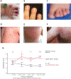
Results: LD was associated with distinctive features, including acute erythematous clubbing and a high frequency of anaphylactic reactions to the interleukin (IL)-6 inhibitor, tocilizumab. Serum ferritin elevation and/or significant lymphopaenia preceded LD detection. The most prevalent chest CT pattern was septal thickening, involving the periphery of multiple lobes ± ground-glass opacities. The predominant pathology (23 of 36) was pulmonary alveolar proteinosis and/or endogenous lipoid pneumonia (PAP/ELP), with atypical features including regional involvement and concomitant vascular changes. Apparent severe delayed drug hypersensitivity occurred in some cases. The 5-year survival was 42%. Whole exome sequencing (20 of 61) did not identify a novel monogenic defect or likely causal PAP-related or macrophage activation syndrome (MAS)-related mutations. Trisomy 21 and young sJIA onset increased LD risk. Exposure to IL-1 and IL-6 inhibitors (46 of 61) was associated with multiple LD features. By several indicators, severity of sJIA was comparable in drug-exposed subjects and published sJIA cohorts. MAS at sJIA onset was increased in the drug-exposed, but was not associated with LD features.
Conclusions: A rare, life-threatening lung disease in sJIA is defined by a constellation of unusual clinical characteristics. The pathology, a PAP/ELP variant, suggests macrophage dysfunction. Inhibitor exposure may promote LD, independent of sJIA severity, in a small subset of treated patients. Treatment/prevention strategies are needed.
Keywords: DMARDs (biologic); adult onset still's disease; inflammation; juvenile idiopathic arthritis; treatment.
© Author(s) (or their employer(s)) 2019. No commercial re-use. See rights and permissions. Published by BMJ.
Conflict of interest statement
Competing interests: VES reports personal fees from Novartis. GD reports personal fees from Novartis. SC reports personal fees from Novartis and grants from AB2 Bio. GS reports personal fees from Novartis. KB reports personal fees from Novartis. RQC is co-PI of an investigator-initiated clinical trial funded by SOBI. RD reports personal fees from Boehringer Ingelheim, other from NowVitals, personal fees and other from Triple Endoscopy, other from Earables, and NowVitals with patents and lung-related device development. AAG reports grants and personal fees from Novartis and grants from NovImmune. SL reports personal fees from Novartis. RS reports personal fees from Novartis, NovImmune and SOBI. SS reports personal fees from Novartis. MLS reports personal fees from Novartis. LRY reports other from Up-To-Date and other from Boehringer Ingelheim, outside the submitted work. EDM reports grants from Novartis.
Figures


Comment in
-
Storm Warning: Lung Disease in Systemic Juvenile Idiopathic Arthritis.Arthritis Rheumatol. 2019 Nov;71(11):1773-1775. doi: 10.1002/art.41071. Epub 2019 Sep 22. Arthritis Rheumatol. 2019. PMID: 31390168 Free PMC article. No abstract available.
-
Response to: 'Effectiveness and safety of ruxolitinib for the treatment of refractory systemic idiopathic juvenile arthritis like associated with interstitial lung disease: case report' by Bader-Meunier et al.Ann Rheum Dis. 2022 Feb;81(2):e21. doi: 10.1136/annrheumdis-2020-217000. Epub 2020 Feb 13. Ann Rheum Dis. 2022. PMID: 32054603 No abstract available.
-
Effectiveness and safety of ruxolitinib for the treatment of refractory systemic idiopathic juvenile arthritis like associated with interstitial lung disease : a case report.Ann Rheum Dis. 2022 Feb;81(2):e20. doi: 10.1136/annrheumdis-2020-216983. Epub 2020 Feb 13. Ann Rheum Dis. 2022. PMID: 32054604 No abstract available.
-
Successful treatment of plasma exchange for refractory systemic juvenile idiopathic arthritis complicated with macrophage activation syndrome and severe lung disease.Ann Rheum Dis. 2022 Apr;81(4):e61. doi: 10.1136/annrheumdis-2020-217390. Epub 2020 Apr 3. Ann Rheum Dis. 2022. PMID: 32245894 No abstract available.
-
Response to: 'Successful treatment of plasma exchange for refractory systemic juvenile idiopathic arthritis complicated with macrophage activation syndrome and severe lung disease' by Sato et al.Ann Rheum Dis. 2022 Apr;81(4):e62. doi: 10.1136/annrheumdis-2020-217426. Epub 2020 Apr 21. Ann Rheum Dis. 2022. PMID: 32317313 No abstract available.
References
-
- Minoia F, Davì S, Horne A, et al. Clinical features, treatment, and outcome of macrophage activation syndrome complicating systemic juvenile idiopathic arthritis: a multinational, multicenter study of 362 patients. Arthritis Rheumatol 2014;66:3160–9. - PubMed
-
- Ruperto N, Brunner HI, Quartier P, et al. Two randomized trials of canakinumab in systemic juvenile idiopathic arthritis. N Engl J Med 2012;367:2396–406. - PubMed
-
- De Benedetti F, Brunner HI, Ruperto N, et al. Randomized trial of tocilizumab in systemic juvenile idiopathic arthritis. N Engl J Med 2012;367:2385–95. - PubMed
Publication types
MeSH terms
Supplementary concepts
Grants and funding
LinkOut - more resources
Full Text Sources
Medical
Research Materials

