. 2019 Nov 1;6(6):ENEURO.0134-19.2019.
doi: 10.1523/ENEURO.0134-19.2019. Print 2019 Nov/Dec.

Mecp2 Deletion from Cholinergic Neurons Selectively Impairs Recognition Memory and Disrupts Cholinergic Modulation of the Perirhinal Cortex

Affiliations

Mecp2 Deletion from Cholinergic Neurons Selectively Impairs Recognition Memory and Disrupts Cholinergic Modulation of the Perirhinal Cortex

Elizabeth C Ballinger et al. eNeuro. .

Abstract

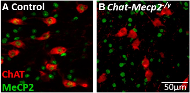
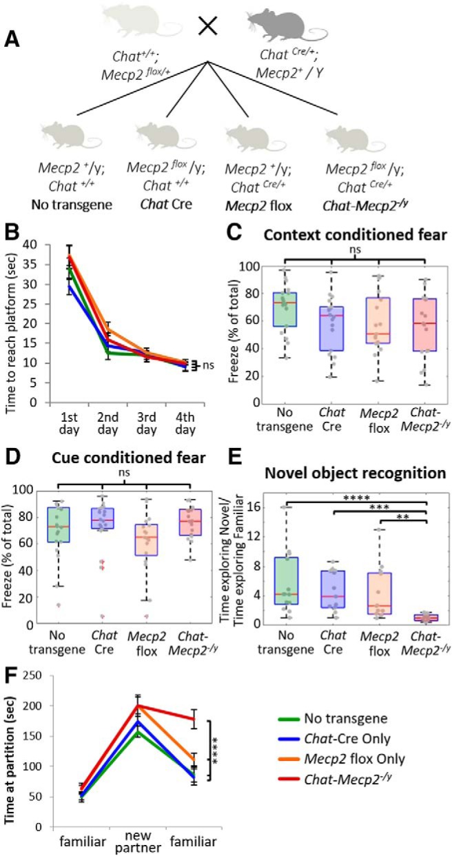
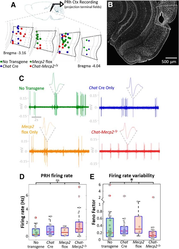
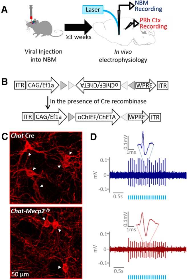
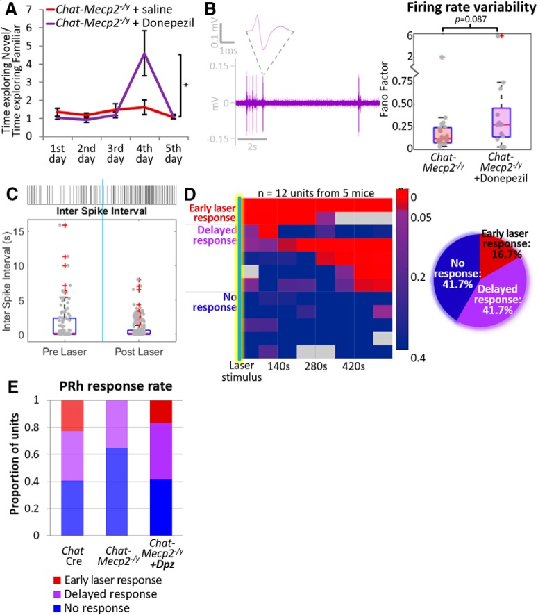
Rett Syndrome is a neurological disorder caused by mutations in the gene encoding methyl CpG binding protein 2 (MeCP2) and characterized by severe intellectual disability. The cholinergic system is a critical modulator of cognitive ability and is affected in patients with Rett Syndrome. To better understand the importance of MeCP2 function in cholinergic neurons, we studied the effect of selective Mecp2 deletion from cholinergic neurons in mice. Mice with Mecp2 deletion from cholinergic neurons were selectively impaired in assays of recognition memory, a cognitive task largely mediated by the perirhinal cortex (PRH). Deletion of Mecp2 from cholinergic neurons resulted in profound alterations in baseline firing of L5/6 neurons and eliminated the responses of these neurons to optogenetic stimulation of cholinergic input to PRH. Both the behavioral and the electrophysiological deficits of cholinergic Mecp2 deletion were rescued by inhibiting ACh breakdown with donepezil treatment.

Keywords: Mecp2; Rett Syndrome; acetylcholine; perirhinal; recognition.


Figures

Figure 1.
Figure 1.
MeCP2 expression is eliminated in cholinergic neurons in Chat-Mecp2-/ymice. A, In a representative slice from the basal forebrain of a no transgene control mouse, MeCP2 expression (green) is apparent in the nuclei of cholinergic neurons (anti-ChAT stained, red). B, In a Chat-Mecp2-/ymouse, however, MeCP2 expression in cholinergic neurons was not detected.
Figure 2.
Figure 2.
Mecp2 cholinergic selective knock-out mice are impaired in recognition memory of either an object or a conspecific. A, Mecp2 flox mice were crossed with Chat Cre mice to generate Mecp2 selective knock-out mice (Chat-Mecp2-/y) and all three genetic controls. A–E, Chat-Mecp2-/y mice (A) performed at control levels on the Morris water maze (B; repeated-measures ANOVA: interaction effect for genotype × testing day: Wilks’ lambda = 0.827, F(9,100) = 0.902, p = 0.527), context-conditioned fear (C; Kruskal–Wallis test: H(3) = 4.64, p = 0.20), and cue-conditioned fear (D; Kruskal–Wallis test: H(3) = 6.46, p = 0.09). E, However, Chat-Mecp2-/y mice were impaired on novel object recognition (Kruskal–Wallis test: H(3) = 22.97, p < 0.0005). Post hoc comparisons revealed that Chat-Mecp2-/y mice were significantly different from all three genetic controls (no transgene, p = 0.0001; Chat Cre, p = 0.0004; Mecp2 flox, p = 0.0079). F, Chat-Mecp2-/y mice showed reduced preference for the novel object introduced on day 4 (novel/familiar object ratio: mean, 1.038; SD, 0.48) than all three genetic controls (no transgene novel/familiar object ratio: mean, 5.6; SD, 4.30; Chat Cre novel/familiar object ratio: mean, 4.63; SD, 2.68; Mecp2 flox novel/familiar object ratio: mean, 4.18; SD, 3.62). Chat-Mecp2-/y mice were also impaired on the partition test (repeated-measures ANOVA: interaction effect for genotype × behavior session: Wilks’ lambda = 0.64; F(6,118) = 4.908; p < 0.0005). Pairwise comparisons revealed that Chat-Mecp2-/y were significantly different from both no transgene mice (p < 0.0005) and Chat Cre mice (p = 0.001), although the difference between Chat-Mecp2-/yand Mecp2 flox mice did not reach significance (p = 0.079). On the partition test, Chat-Mecp2-/y mice were impaired in their ability to recognize a familiar mouse and spent longer interacting with the familiar mouse on re-presentation (mean, 178.05 s; SD, 63.02) than any of the genetic controls (no transgene: mean, 86.58 s; SD, 47.92; Chat Cre: mean, 82.38 s; SD, 51.89; Mecp2 flox: mean, 111.31s; SD, 43.97). Error bars represent the SEM. **p ≤ 0.01, ***p ≤ 0.001, ****p ≤ 0.0005, ns = non significant.
Figure 3.
Figure 3.
Neuronal firing in the PRH is highly variable, and this variability is lost in Chat-Mecp2-/y mice. A, In vivo recordings were collected from layers 5 and 6 of the PRH. B, Sample Nissl staining and electrolytic lesion marking recording sites in the PRH (white dotted line). C, Representative recordings show the highly variable baseline firing in controls that is lost in Chat-Mecp2-/y mice. D, There was no difference between genotypes in baseline firing rates in the PRH (Kruskal–Wallis test: H(3) = 6.62; p = 0.085). E, Variability of firing rate as measured by the Fano factor was significantly different between groups (Kruskal–Wallis test: H(3) = 8.92; p = 0.03). Chat-Mecp2-/y mice had a lower firing rate variability than all three controls (no transgene: mean FF = 0.612 ± 1.250; Chat Cre: mean FF = 0.474 ± 0.650; Mecp2 flox: mean FF = 0.447 ± 0.365; Chat-Mecp2-/y: mean FF = 0.248 ± 0.431). No transgene: n = 24 units from 6 mice; Chat Cre: n = 22 units from 11 mice; Mecp2 flox: n = 6 units from 3 mice; Chat-Mecp2-/y: n = 20 units from 8 mice. *p ≤ 0.05, ns = non significant.
Figure 4.
Figure 4.
Strategy for optogenetic stimulation of cholinergic neurons. A, Schematic of the experimental paradigm. B, A viral vector encoding an optically activated excitatory ion channel is injected into the NBM. The viral vector is of a flip excision switch design such that it will be expressed only in the presence of Cre recombinase. C, Representative images of virally labeled cholinergic neurons (white arrowheads) from a control mouse (blue, top) and a Chat-Mecp2-/y mouse (red, bottom). D, Representative optically evoked action potentials in the NBM of a control mouse (top) and a Chat-Mecp2-/y mouse (bottom). The timing of laser pulses delivered into the NBM is indicated by light blue hash marks.
Figure 5.
Figure 5.
PRH response to the stimulation of endogenous cholinergic signaling is impaired in Chat-Mecp2-/y mice. A, B, Representative data from a PRH unit in a control mouse exhibiting a response to stimulation of cholinergic input (A) and a PRH unit from an Chat-Mecp2-/y mouse (B). Top, Representative raster plot of spikes. Vertical light blue bar indicates timing of optical stimulation. Bottom, Box plot of interspike intervals. C, Heat map of p values as a function of time since optical stimulation for PRH units from control mice (left) and from Chat-Mecp2-/y mice (right). Responses either occurred in the first time period following laser stimulation or were delayed. Each row represents a separate unit. The results are summarized in pie charts at bottom. D, Summary of differing response rates between control and Chat-Mecp2-/y units (χ2 test for homogeneity: CR(2) = 6.02; p = 0.049). *p ≤ 0.05.
Figure 6.
Figure 6.
Donepezil treatment of Chat-Mecp2-/y mice rescues both behavioral and electrophysiological impairments. A, Chronic treatment with systemic donepezil, a drug that inhibits acetylcholinesterase, administered subcutaneously for 2 weeks rescued behavioral impairment (Chat-Mecp2-/y+ saline, n = 8; Chat-Mecp2-/y + Dpz, n = 10; Wilcoxon rank sum test: rank sum = 120; p = 0.03). B, Representative raw data trace showing that baseline firing variability was rescued on treatment with donepezil. Firing rate variability was increased in Chat-Mecp2-/y mice after treatment with donepezil (Chat-Mecp2-/y, mean FF = 0.248 ± 0.431; Chat-Mecp2-/y+ Dpz, mean FF = 0.735 ± 1.620), although this difference did not reach statistical significance (Wilcoxon rank sum test: rank sum = 293, p = 0.087). C, Sample response to optogenetic stimulation in a Chat-Mecp2-/y mouse after treatment with donepezil. At top is shown a raster plot of action potentials before and after stimulation of cholinergic input (indicated by light blue vertical bar). At bottom is shown a box plot of interspike intervals obtained before and after optical stimulation of cholinergic neurons in the NBM. D, Heat map of p values as a function of time since optical stimulation. Responses either occurred in the first time period following laser stimulation or were delayed and are summarized in the pie chart at bottom. E, Summary of response types. Early laser responses were restored in Chat-Mecp2-/y mice treated with donepezil. (χ2 test for homogeneity: CR(2) = 4.15; p = 0.126). *p ≤ 0.05.

Similar articles

Cited by

References

    1. Allen TG, Abogadie FC, Brown DA (2006) Simultaneous release of glutamate and acetylcholine from single magnocellular “cholinergic” basal forebrain neurons. J Neurosci 26:1588–1595. 10.1523/JNEUROSCI.3979-05.2006 - DOI - PMC - PubMed
    1. Amir RE, Van den Veyver IB, Wan M, Tran CQ, Francke U, Zoghbi HY (1999) Rett syndrome is caused by mutations in X-linked MECP2, encoding methyl-CpG-binding protein 2. Nat Genet 23:185–188. 10.1038/13810 - DOI - PubMed
    1. Ballinger EC, Ananth M, Talmage DA, Role LW (2016) Basal forebrain cholinergic circuits and signaling in cognition and cognitive decline. Neuron 91:1199–1218. 10.1016/j.neuron.2016.09.006 - DOI - PMC - PubMed
    1. Beaudin SA, Singh T, Agster KL, Burwell RD (2013) Borders and comparative cytoarchitecture of the perirhinal and postrhinal cortices in an F1 hybrid mouse. Cereb Cortex 23:460–476. 10.1093/cercor/bhs038 - DOI - PMC - PubMed
    1. Brašic´ JR, Bibat G, Kumar A, Zhou Y, Hilton J, Yablonski ME, Dogan AS, Guevara MR, Stephane M, Johnston M, Wong DF, Naidu S (2012) Correlation of the vesicular acetylcholine transporter densities in the striata to the clinical abilities of women with rett syndrome. Synapse 66:471–482. 10.1002/syn.21515 - DOI - PMC - PubMed

Publication types

MeSH terms

LinkOut - more resources