. 2020 Jan;61(1):144-151.
doi: 10.2967/jnumed.119.231845. Epub 2019 Sep 27.

PennPET Explorer: Human Imaging on a Whole-Body Imager

Affiliations

PennPET Explorer: Human Imaging on a Whole-Body Imager

Austin R Pantel et al. J Nucl Med. 2020 Jan.

Abstract

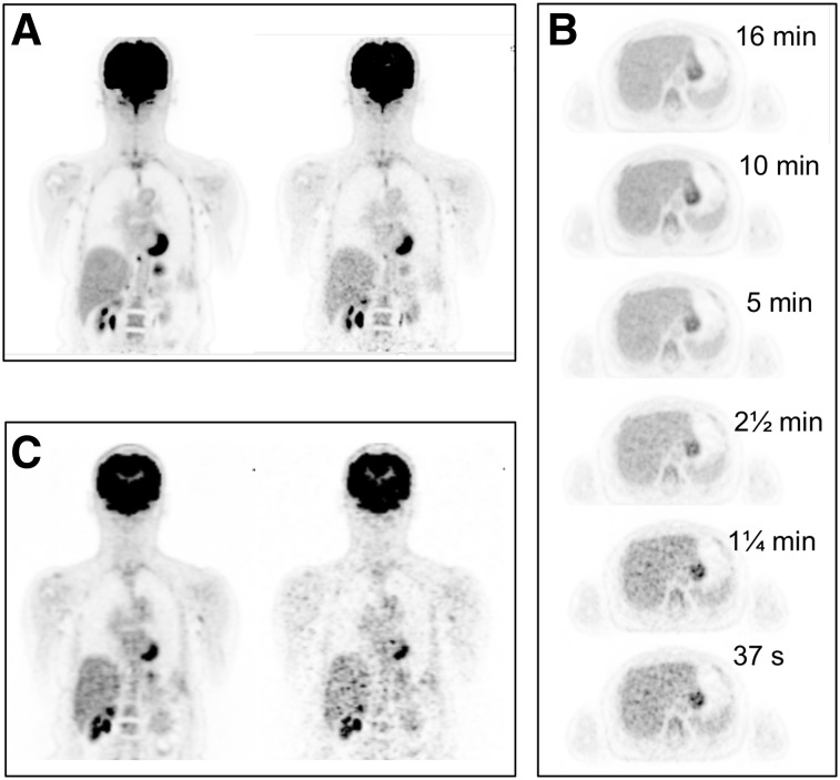
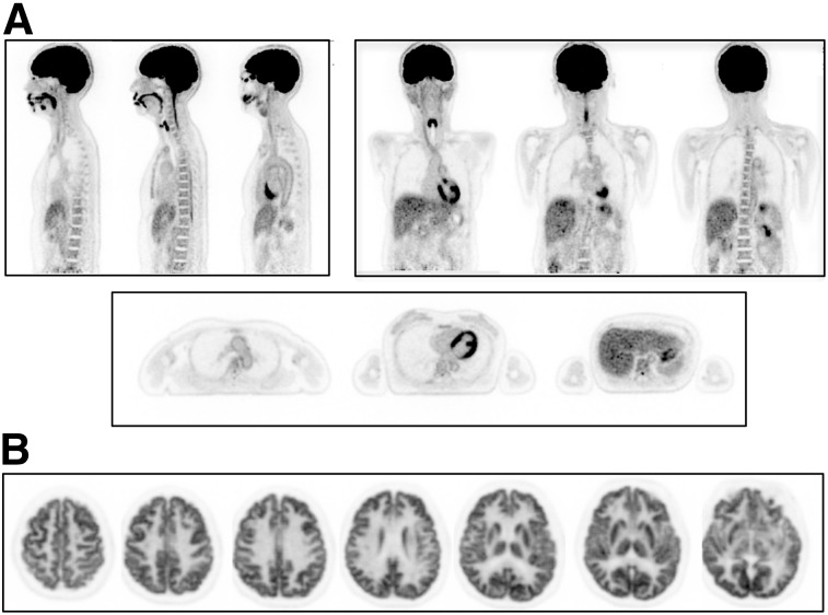
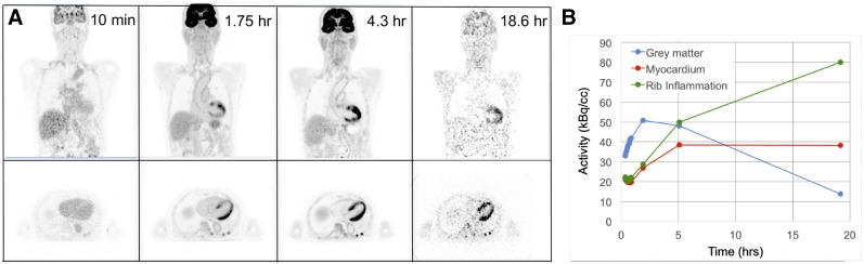
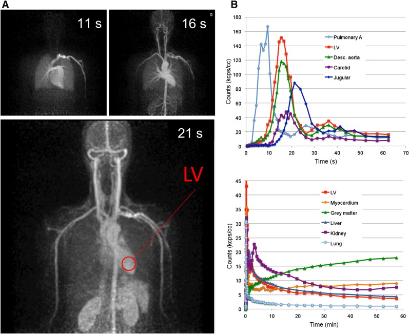
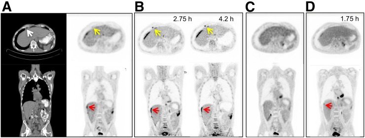
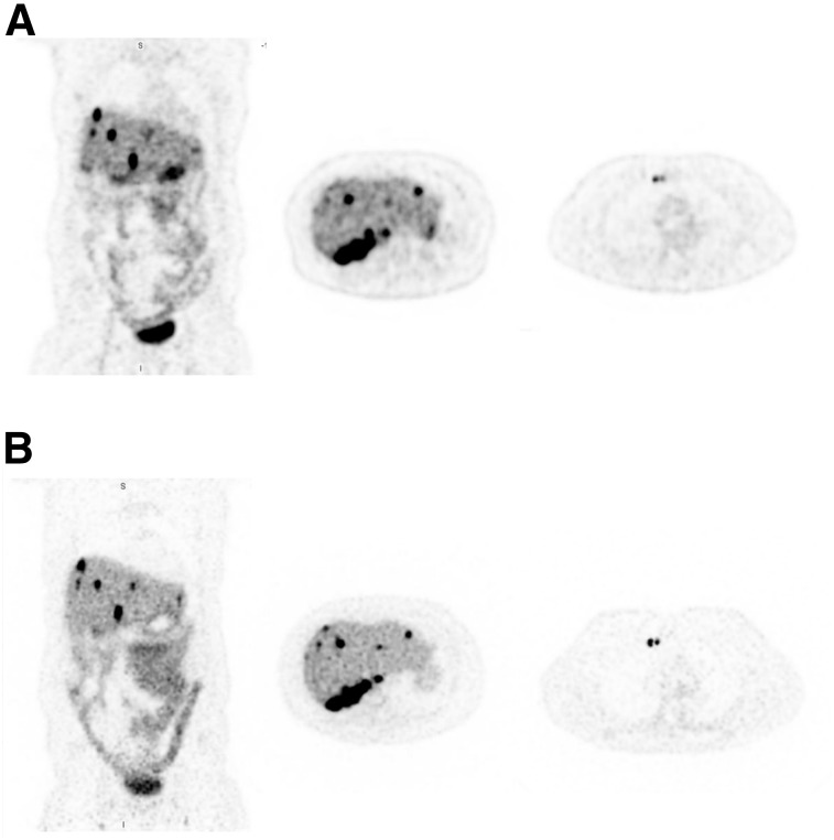
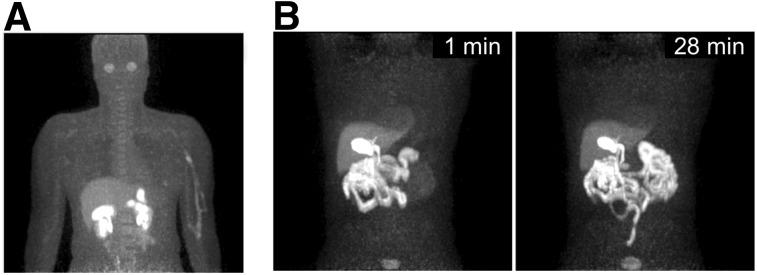
The PennPET Explorer, a prototype whole-body imager currently operating with a 64-cm axial field of view, can image the major body organs simultaneously with higher sensitivity than that of commercial devices. We report here the initial human imaging studies on the PennPET Explorer, with each study designed to test specific capabilities of the device. Methods: Healthy subjects were imaged with FDG on the PennPET Explorer. Subsequently, clinical subjects with disease were imaged with 18F-FDG and 68Ga-DOTATATE, and research subjects were imaged with experimental radiotracers. Results: We demonstrated the ability to scan for a shorter duration or, alternatively, with less activity, without a compromise in image quality. Delayed images, up to 10 half-lives with 18F-FDG, revealed biologic insight and supported the ability to track biologic processes over time. In a clinical subject, the PennPET Explorer better delineated the extent of 18F-FDG-avid disease. In a second clinical study with 68Ga-DOTATATE, we demonstrated comparable diagnostic image quality between the PennPET scan and the clinical scan, but with one fifth the activity. Dynamic imaging studies captured relatively noise-free input functions for kinetic modeling approaches. Additional studies with experimental research radiotracers illustrated the benefits from the combination of large axial coverage and high sensitivity. Conclusion: These studies provided a proof of concept for many proposed applications for a PET scanner with a long axial field of view.

Keywords: PET; human imaging; whole-body imager.


Figures

FIGURE 1.
FIGURE 1.
(A) 18F-FDG PET coronal images of subject 1 on PennPET acquired at 1.5 h after injection of 18F-FDG for 16 min (left) and 2 min (right). (B) Transverse images of liver from PennPET over range of scan durations. (C) Coronal images from SOC clinical PET acquired at 0.75 h after injection for 16 min (left) and 2 min (right).
FIGURE 2.
FIGURE 2.
(A) 18F-FDG PET images of subject 2 (sagittal, coronal, and axial) on PennPET (10-min scan). (B) Transverse images on PennPET after subject was moved so that brain was positioned in center of axial FOV (10-min scan).
FIGURE 3.
FIGURE 3.
(A) 18F-FDG PET coronal images of subject 3 acquired at 4 time points after injection. First time point is 3-min scan; other time points are as noted in Table 1. (B) Time–activity curves for brain, myocardium, and rib fracture from same subject. Plotted points are at mid time of each scan.
FIGURE 4.
FIGURE 4.
(A) 18F-FDG PET maximum-intensity projections of subject 7, each 1 s in duration, at 3 time points from dynamic scan. (B) Time–activity curves of blood input function measured at several vessels over first minute after injection, and time–activity curves of major organs over first hour after injection. LV = left ventricle.
FIGURE 5.
FIGURE 5.
Clinical 18F-FDG PET/CT images (transverse and coronal) from subject 5, with metastatic colon cancer, acquired with standard clinical protocol. (B) PennPET image acquired 2.75 and 4.2 h after injection (10-min scans). Matched coronal and transverse slices are shown. Red arrows denote perihepatic disease; yellow arrows denote epiphrenic lymph node. (C) Follow-up clinical scan at 3 mo (subject 10). (D) Corresponding PennPET image (20-min scan) demonstrating improvement in perihepatic disease and epiphrenic lymph node.
FIGURE 6.
FIGURE 6.
(A) SOC 68Ga-DOTATATE PET images (coronal and transverse) of subject 8, with metastatic neuroendocrine tumor. (B) Coronal and transverse images from same subject on PennPET acquired 3.5 h after injection (20-min scan).
FIGURE 7.
FIGURE 7.
(A) Maximum-intensity projection (3-min scan) of 18F-labeled NOS PET (subject 6). (B) Maximum-intensity projections of 18F-fluortriopride PET (subject 9) for 1-min duration shown at 1 min (left) and 28 min (right) after drinking Ensure (Abbott Laboratories) to stimulate emptying of radiotracer from gallbladder.

References

    1. Hsu DF, Ilan E, Peterson WT, Uribe J, Lubberink M, Levin CS. Studies of a next-generation silicon-photomultiplier–based time-of-flight PET/CT system. J Nucl Med. 2017;58:1511–1518. - PubMed
    1. Reddin JS, Scheuermann JS, Bharkhada D, et al. Performance evaluation of the SiPM-based Siemens Biograph Vision PET/CT system. In: 2018 IEEE Nuclear Science Symposium and Medical Imaging Conference Proceedings (NSS/MIC). Piscataway, NJ: IEEE; 2018.
    1. Rausch I, Ruiz A, Valverde-Pascual I, Cal-González J, Beyer T, Carrio I. Performance evaluation of the Vereos PET/CT system according to the NEMA NU2-2012 standard. J Nucl Med. 2019;60:561–567. - PubMed
    1. Cherry SR, Jones T, Karp JS, Qi J, Moses WW, Badawi RD. Total-body PET: maximizing sensitivity to create new opportunities for clinical research and patient care. J Nucl Med. 2018;59:3–12. - PMC - PubMed
    1. Cherry SR, Badawi RD, Karp JS, Moses WW, Price P, Jones T. Total-body imaging: transforming the role of positron emission tomography. Sci Transl Med. 2017;9:eaaf6169. - PMC - PubMed

Publication types

Substances

LinkOut - more resources