. 2019 Nov 8;294(45):17155-17165.
doi: 10.1074/jbc.RA119.010271. Epub 2019 Sep 27.

Atomic-resolution crystal structures of the immune protein conglutinin from cow reveal specific interactions of its binding site with N-acetylglucosamine

Affiliations

Atomic-resolution crystal structures of the immune protein conglutinin from cow reveal specific interactions of its binding site with N-acetylglucosamine

Janet M Paterson et al. J Biol Chem. .

Abstract

Bovine conglutinin is an immune protein that is involved in host resistance to microbes and parasites and interacts with complement component iC3b, agglutinates erythrocytes, and neutralizes influenza A virus. Here, we determined the high-resolution (0.97-1.46 Å) crystal structures with and without bound ligand of a recombinant fragment of conglutinin's C-terminal carbohydrate-recognition domain (CRD). The structures disclosed that the high-affinity ligand N-acetyl-d-glucosamine (GlcNAc) binds in the collectin CRD calcium site by interacting with the O3' and O4' hydroxyls alongside additional specific interactions of the N-acetyl group oxygen and nitrogen with Lys-343 and Asp-320, respectively. These residues, unique to conglutinin and differing both in sequence and in location from those in other collectins, result in specific, high-affinity binding for GlcNAc. The binding pocket flanking residue Val-339, unlike the equivalent Arg-343 in the homologous human surfactant protein D, is sufficiently small to allow conglutinin Lys-343 access to the bound ligand, whereas Asp-320 lies in an extended loop proximal to the ligand-binding site and bounded at both ends by conserved residues that coordinate to both calcium and ligand. This loop becomes ordered on ligand binding. The electron density revealed both α and β anomers of GlcNAc, consistent with the added α/βGlcNAc mixture. Crystals soaked with α1-2 mannobiose, a putative component of iC3b, reported to bind to conglutinin, failed to reveal bound ligand, suggesting a requirement for presentation of mannobiose as part of an extended physiological ligand. These results reveal a highly specific GlcNAc-binding pocket in conglutinin and a novel collectin mode of carbohydrate recognition.

Keywords: carbohydrate-binding protein; collectin; complement; conglutinin; crystal structure; host-pathogen interaction; innate immunity; lectin; structural biology; surfactant protein.


Conflict of interest statement

The authors declare that they have no conflicts of interest with the contents of this article

Figures
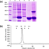
Figure 1.
Figure 1.
Expression and purification of rfBC. a, reducing 15% SDS-PAGE analysis of the expression of rfBC. Lane 1, low molecular weight markers; lane 2, total cell lysate of uninduced cells; lane 3, total cell lysate of induced cells harvested 2 h post induction and chilled for 30 min on ice with shaking; lane 4, sonicated supernatant; lane 5, refolded protein after overnight dialysis; lane 6, dialysate pellet containing insoluble material. b, gel-filtration chromatography elution profile of rfBC. Following ion exchange purification of the expressed rfBC protein (Q-Sepharose followed by Mono Q) the purified protein was run at a flow rate of 1 ml/min (tris/EDTA buffer) through a Superdex75 gel-filtration column. 0.5 ml volume fractions were collected. The molecular mass standards alcohol dehydrogenase (150 kDa), BSA (66 kDa), carbonic anhydrase (29 kDa), and cytochrome C (14.4 kDa) used for calibration are shown.
Figure 2.
Figure 2.
Sequence of the rfBC recombinant fragment. The first five amino acids GSAEA (GLAEV in conglutinin) are vector derived. The truncated monomeric fragment used for structure determination is underlined with the sites of proteolysis within the α helical coiled coil neck region, leading to the truncated fragment, indicated by the vertical arrows.
Figure 3.
Figure 3.
Electron density for the αGlcNAc-1-P ligand bound to conglutinin. Conglutinin ligand-binding site with αGlcNAc-1-P ligand shown as ball-and-stick model. The 1.0 Å resolution electron density map (blue) is 2mFo − DFc contoured at 1σ. The calcium ion is in green with calcium and ligand coordinating residues Glu-315, Asn-317, Glu-325, and Asn-337 are shown as stick models.
Figure 4.
Figure 4.
Structure of the CRD. Overlay of conglutinin (gray), hSP-D (blue), and pSP-D (orange) CRDs generated by superposing (least squares fit of main chain) conglutinin residues 232–319 and 325–351 with equivalent residues in PDB ID 1PWB and PDB ID 4DN8. a, overlay illustrating the location of the calcium ions, Ca1, Ca2, and Ca3, with Ca3 only found in hSP-D. The long loop indicated corresponds to residues 318–324 in conglutinin. b, GlcNAc-bound conglutinin structure with ligand coordinating residues Asp-320, from the long loop, and Lys-343 shown as sticks. For clarity only βGlcNAc and conglutinin Ca1 are shown.
Figure 5.
Figure 5.
GlcNAc binding in conglutinin CRD ligand site showing key amino acids and interactions between bound ligand and protein. Both αGlcNAc and βGlcNAc are seen in the binding site with the anomeric carbon C1 indicated. Amino acid interactions are shown for αGlcNAc. βGlcNAc interactions are the same except for that with Glu-321 which is only present for αGlcNAc. The Lys-246 interacting residue from a neighboring subunit via a crystal contact is shown in blue and calcium ion Ca1 is shown as a green sphere.
Figure 6.
Figure 6.
Sequence alignment in the calcium- and ligand-binding region in the collectin CRD. Conserved residues are shaded gray and conserved Ca1 coordinators are yellow. The specific GlcNAc conglutinin N-acetyl interacting residues Asp-320 and Lys-343 along with the binding site flanking residues of hSP-D, Asp-325, and Arg-343 (see Shrive et al., 32) are highlighted in cyan.
Figure 7.
Figure 7.
Extended binding site interactions. Conglutinin CRD (surface view) with residues in the extended ligand-binding pocket highlighted in gray with black labels. Corresponding residues in hSP-D (least squares fit of 1PWB CRD to main chain conglutinin residues 232–319 and 325–351) are shown in yellow with orange labels.

References

    1. Holmskov U. L. (2000) Collectins and collectin receptors in innate immunity. APMIS Suppl. 100, 1–59 - PubMed
    1. Crouch E. C. (1998) Structure, biologic properties, and expression of surfactant protein D (SP-D). Biochim. Biophys. Acta 1408, 278–289 10.1016/S0925-4439(98)00073-8 - DOI - PubMed
    1. Lawson P. R., and Reid K. B. M. (2000) The roles of surfactant proteins A and D in innate immunity. Immunol. Rev. 173, 66–78 10.1034/j.1600-065X.2000.917308.x - DOI - PubMed
    1. Sano H., and Kuroki Y. (2005) The lung collectins, SP-A and SP-D, modulate pulmonary innate immunity. Mol. Immunol. 42, 279–287 10.1016/j.molimm.2004.07.014 - DOI - PubMed
    1. Thiel S. (1992) Mannan-binding protein, a complement activating animal lectin. Immunopharmacology 24, 91–99 10.1016/0162-3109(92)90015-5 - DOI - PubMed

Publication types

LinkOut - more resources