Review
. 2019 Sep 27;12(10):dmm040691.
doi: 10.1242/dmm.040691.

Model organisms at the heart of regeneration

Affiliations
Review

Model organisms at the heart of regeneration

Eleanor L Price et al. Dis Model Mech. .

Abstract

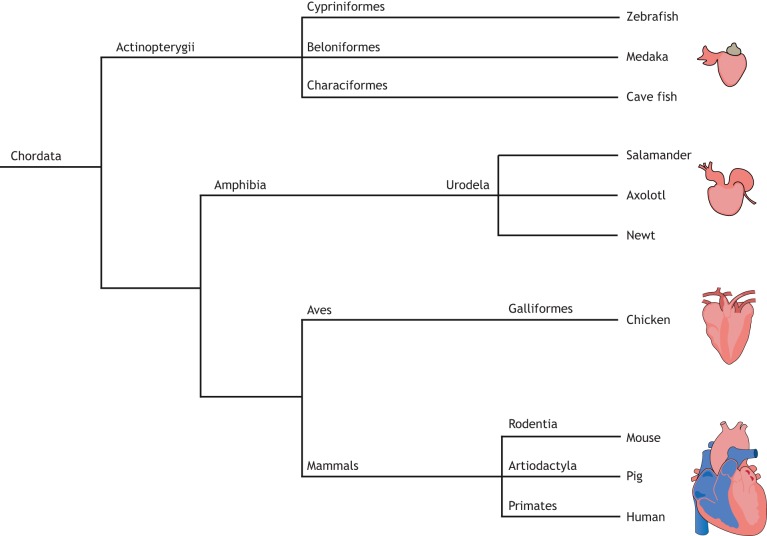
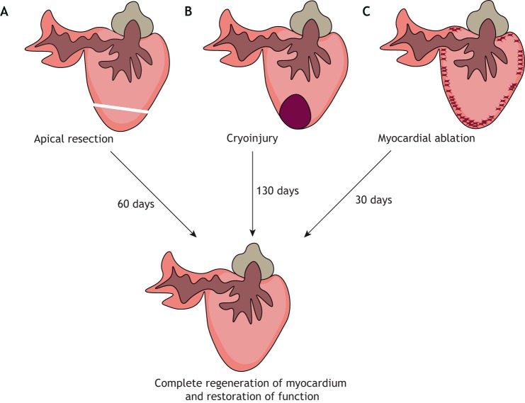
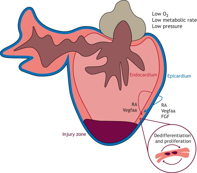
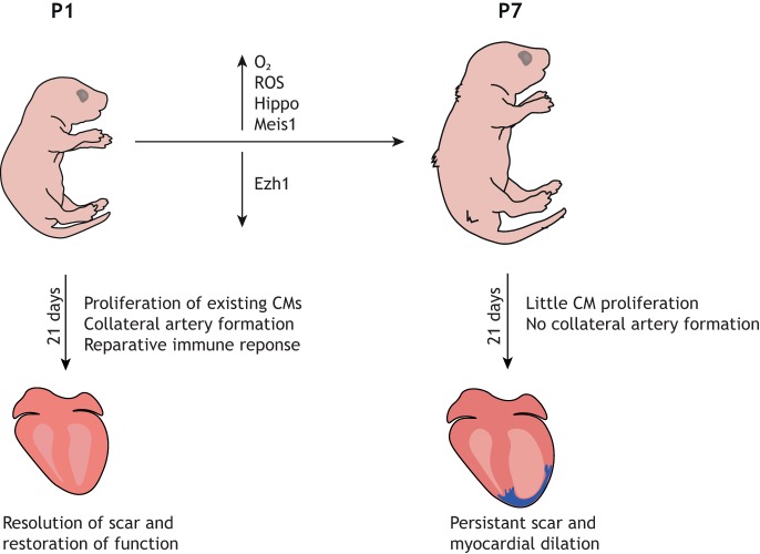
Heart failure is a major cause of death worldwide owing to the inability of the adult human heart to regenerate after a heart attack. However, many vertebrate species are capable of complete cardiac regeneration following injury. In this Review, we discuss the various model organisms of cardiac regeneration, and outline what they have taught us thus far about the cellular and molecular responses essential for optimal cardiac repair. We compare across different species, highlighting evolutionarily conserved mechanisms of regeneration and demonstrating the importance of developmental gene expression programmes, plasticity of the heart and the pathophysiological environment for the regenerative response. Additionally, we discuss how the findings from these studies have led to improvements in cardiac repair in preclinical models such as adult mice and pigs, and discuss the potential to translate these findings into therapeutic approaches for human patients following myocardial infarction.

Keywords: Cardiac regeneration; Cardiac repair; Cardiomyocytes; Myocardial infarction; Regenerative medicine.


Conflict of interest statement

Competing interestsThe authors declare no competing or financial interests.

Figures

Fig. 1.
Fig. 1.
Animal models of cardiac regeneration. Phylogenetic tree to demonstrate the evolutionary divergence of the different vertebrate species used in the study of cardiac regeneration.
Fig. 2.
Fig. 2.
Methods of cardiac injury in the zebrafish. (A) Apical resection involves surgical removal of ∼20% of the ventricular apex. (B) In the cryoinjury model, ∼25% of the ventricle is damaged by placing a cryoprobe onto the heart. (C) Genetic ablation leads to the loss of ∼60% of cardiomyocytes.
Fig. 3.
Fig. 3.
Cardiac regeneration in zebrafish. Following cardiac injury, diploid cardiomyocytes undergo dedifferentiation and proliferate to replace the damaged tissue. Signals from the activated epicardium and endocardium, such as retinoic acid (RA) and vascular endothelial growth factor Aa (Vegfaa), stimulate such proliferation, whilst epicardial fibroblast growth factor (FGF) signalling induces neovascularisation to restore blood flow.
Fig. 4.
Fig. 4.
Mouse models of neonatal cardiac regeneration. Following a thoracotomy, the heart is exposed from the chest and can be injured by either: (A) removal of the ventricular apex; (B) ligation of the left anterior descending coronary artery (LAD) using a suture; or (C) a cryoprobe held against the ventricle wall.
Fig. 5.
Fig. 5.
Loss of cardiac regeneration potential in neonatal mice. Within the first week of life, the neonatal mouse loses the ability to regenerate its heart. This diagram highlights some of the key changes that occur within the first week of life that contribute to the loss of regenerative capacity. CM, cardiomyocyte; ROS, reactive oxygen species.

References

    1. Ai S., Yu X., Li Y., Peng Y., Li C., Yue Y., Tao G., Li C., Pu W. T. and He A. (2017). Divergent requirements for EZH1 in heart development versus regeneration. Circ. Res. 121, 106-112. 10.1161/CIRCRESAHA.117.311212 - DOI - PMC - PubMed
    1. Andersen D. C., Ganesalingam S., Jensen C. H. and Sheikh S. P. (2014). Do neonatal mouse hearts regenerate following heart apex resection? Stem Cell Reports 2, 406-413. 10.1016/j.stemcr.2014.02.008 - DOI - PMC - PubMed
    1. Andersen D. C., Jensen C. H., Baun C., Hvidsten S., Zebrowski D. C., Engel F. B. and Sheikh S. P. (2016). Persistent scarring and dilated cardiomyopathy suggest incomplete regeneration of the apex resected neonatal mouse myocardium—A 180 days follow up study. J. Mol. Cell. Cardiol. 90, 47-52. 10.1016/j.yjmcc.2015.11.031 - DOI - PubMed
    1. Arenas Gómez C. M., Gómez Molina A., Zapata J. D. and Delgado J. P. (2017). Limb regeneration in a direct-developing terrestrial salamander, Bolitoglossa ramosi (Caudata: Plethodontidae): limb regeneration in plethodontid salamanders. Regeneration 4, 227-235. 10.1002/reg2.93 - DOI - PMC - PubMed
    1. Arós F., Loma-Osorio A., Vila J., López-Bescós L., Cuñat J., Rodríguez E., San José J. M., Heras M. and Marrugat J. (2006). [Effect of combined beta-blocker and angiotensin-converting enzyme inhibitor treatment on 1-year survival after acute myocardial infarction: findings of the PRIAMHO-II registry]. Rev. Esp. Cardiol. 59, 313-320. 10.1157/13087059 - DOI - PubMed

Publication types