. 2020 Apr;27(4):1383-1397.
doi: 10.1038/s41418-019-0424-4. Epub 2019 Sep 27.

RBX1 prompts degradation of EXO1 to limit the homologous recombination pathway of DNA double-strand break repair in G1 phase

Affiliations

RBX1 prompts degradation of EXO1 to limit the homologous recombination pathway of DNA double-strand break repair in G1 phase

Ying Xie et al. Cell Death Differ. 2020 Apr.

Abstract

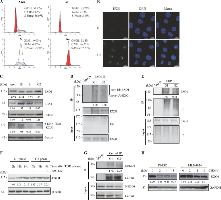
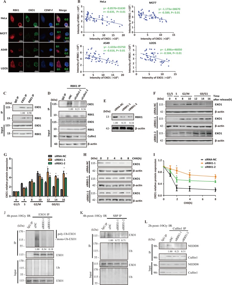
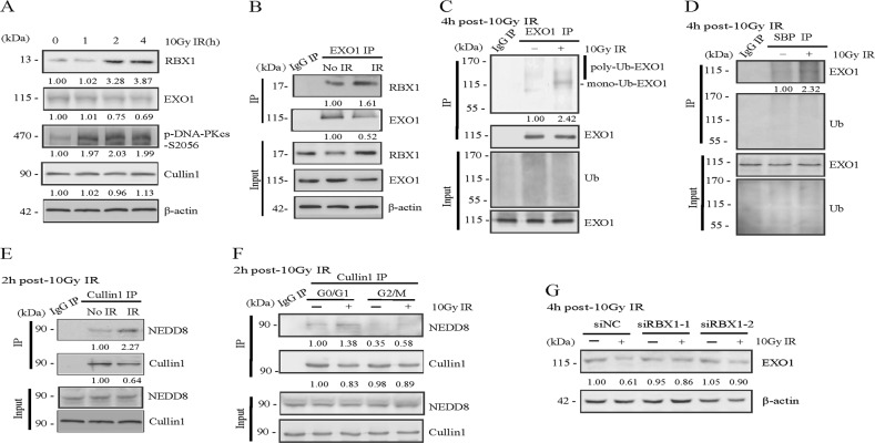
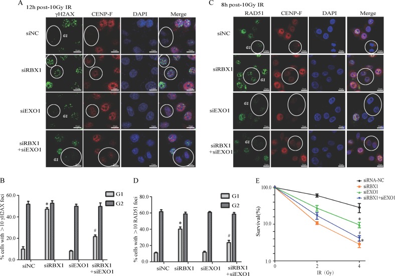
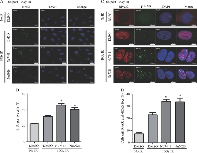
End resection of DNA double-strand breaks (DSBs) to form 3' single-strand DNA (ssDNA) is critical to initiate the homologous recombination (HR) pathway of DSB repair. HR pathway is strictly limited in the G1-phase cells because of lack of homologous DNA as the templates. Exonuclease 1 (EXO1) is the key molecule responsible for 3' ssDNA formation of DSB end resection. We revealed that EXO1 is inactivated in G1-phase cells via ubiquitination-mediated degradation, resulting from an elevated expression level of RING-box protein 1 (RBX1) in G1 phase. The increased RBX1 significantly prompted the neddylation of Cullin1 and contributed to the G1 phase-specific degradation of EXO1. Knockdown of RBX1 remarkedly attenuated the degradation of EXO1 and increased the end resection and HR activity in γ-irradiated G1-phase cells, as demonstrated by the increased formation of RPA32, BrdU, and RAD51 foci. And EXO1 depletion mitigated DNA repair defects due to RBX1 reduction. Moreover, increased autophosphorylation of DNA-PKcs at S2056 was found to be responsible for the higher expression level of the RBX1 in the G1 phase. Inactivation of DNA-PKcs decreased RBX1 expression, and simultaneously increased EXO1 expression and DSB end resection in G1-phase cells. This study demonstrates a new mechanism for restraining the HR pathway of DNA DSB repair in G1 phase via RBX1-prompted inactivation of EXO1.


Conflict of interest statement

The authors declare that they have no conflict of interest.

Figures

Fig. 1
Fig. 1
High-level ubiquitination mediated suppression of EXO1 in G1-phase cells. a HeLa cells were synchronized to different phases of the cell cycle by double-blockage of thymidine, and the cell phase distributions were monitored by flow cytometry analysis. b EXO1 protein was detected in the synchronized G1- and G2-phase HeLa cells by microscopic observation of immunofluorescent staining with anti-EXO1 antibody. Nuclei were stained with DAPI. c The expression levels of EXO1, RBX1, Cullin1, and phosphorylated DNA-PKcs-S2056 were assessed by western blotting analysis. Among them, phosphorylation of DNA-PKcs, is indicative of cells in different phase of cell cycle. d, e Ubiquitination of EXO1 protein was detected respectively in the synchronized G1- and G2-phase cells. After HeLa were transfected with HA-Ub and SBP-Ub plasmid for 48 h, the immunoprecipitation (IP) product obtained with the EXO1 antibody and S-bead agarose, were subjected to western blotting analysis with the anti-flag antibody and anti-EXO1 antibody separately. In order to achieve the same content of EXO1 for immunoprecipitation, when HA-Ub and SBP-Ub transfected cells were synchronized in G1- and G2-phase using a double thymidine block, cell were pre-treated with proteasome inhibitor MG132 for 2 h. f Hela were arrested at the G1/S boundary followed by a release into fresh media to allow cells to progress through the cell cycle. Cells were synchronized in G2 phase (7–9h) and G1 phase (12–20h) after TDR release. 10 μM MG132 was added in G1 and G2 cells for 2 h and EXO1 contents were detected. g Neddylation of Cullin1 protein was detected separately in the synchronized G1- and G2-phase cells. The immunoprecipitation (IP) product obtained with the Cullin1 antibody was subjected to western blotting analysis with anti-NEDD8 antibody. h The effect of neddylation on the stability of the EXO1 protein. Cells were pre-treated with the neddylation inhibitor MLN4924 or DMSO as a control for 8 h and then co-cultured with cycloheximide (CHX). EXO1 levels were assessed by western blotting analysis at the indicated times after CHX treatment
Fig. 2
Fig. 2
RBX1 mediates the degradation of EXO1 in G1-phase cells by promoting the neddylation of Cullin1 and activating SCF E3 ligase. a Immunofluorescent staining images displayed the negative correlation between RBX1 and EXO1 expression in various types of cells. HeLa, MCF7, A549, and U2OS cells were immunostained with anti-RBX1 antibody (red), anti-EXO1 antibody (green), and anti-CENP-F antibody (blue), and the images were collected under laser confocal microscopy. CENP-F staining was used to distinguish G2/M-phase cells from G1 cells. b Quantitative analysis of the correlation between RBX1 and EXO1 expression in multiple cell lines. The fluorescence intensities of RBX1 and EXO1 were measured using ImageJ software, and a correlation analysis was performed. p value less than 0.05 indicates a significant relationship between RBX1 and EXO1 expression. c The interaction between EXO1 and RBX1 were observed by IP-Western. d After knockdown of Cullin1 by siRNA in HeLa cells, the interaction between EXO1 and RBX1 were assessed. e Western blotting analysis verified the knockdown of RBX1 by siRNA in HeLa cells. f Knockdown of RBX1 augmented the EXO1 protein in G1-phase cells. HeLa cells were depleted of endogenous RBX1 using siRNA and synchronized with double-blockage of thymidine. Then, EXO1 protein levels were detected by western blotting analysis at different times after release from thymidine blockage, and the synchronized cells were monitored by flow cytometry. g Densitometric quantitation of western blotting analysis of EXO1 protein expression. Data are the mean ± standard deviation from three independent experiments. *p< 0.05 compared with the cells treated with the control siRNA-NC. h The effect of knocking down RBX1 on the stability of the EXO1 protein. The RBX1-knockdown cells by specific siRNA and the control cells were cultured with cycloheximide (CHX). EXO1 levels were assessed by western blotting analysis at the indicated times after CHX treatment. i Densitometric quantitation of western blotting analysis of EXO1 protein expression at the indicated times after CHX treatment. Data are the mean ± standard deviation from three independent experiments. *p< 0.05 compared with the cells treated with the control siRNA-NC. j, k Knockdown of RBX1 led to decreased ubiquitination of the EXO1 protein. HeLa cells were co-transfected with siRNA RBX1 and HA-Ub or SBP-Ub plasmid, and the immunoprecipitation (IP) product obtained with the EXO1 antibody and S-bead agarose, were subjected to western blotting analysis with the anti-flag antibody and anti-EXO1 antibody separately. l Knockdown of RBX1 led to decreased neddylation of Cullin1 protein. The immunoprecipitation (IP) production of Cullin1 antibody from the siRNA-mediated RBX1-knockdown HeLa cells and the control cells were subjected to western blotting analysis with anti-NEDD8 antibody
Fig. 3
Fig. 3
Depression of RBX1 causes radiosensitization and deficiency in DNA double-strand break repair. a Cell survival of HeLa cells irradiated with γ-rays by colony-forming ability assay. b Neutral comet assay of radiation-induced DNA double-strand break (DSB) repair in HeLa cells treated with RBX1 siRNA or control siRNA-NC. DNA damage was expressed as the Olive tail moment. c Detection of radiation-induced DNA DSBs by γH2AX foci of immunofluorescent staining in HeLa cells treated with RBX1 siRNA or control siRNA-NC. Quantitative determination of γH2AX foci as DSB indicators in RBX1-knockdown HeLa cells and control cells irradiated with 2 Gy (d) or 10 Gy (e) of γ-rays. In total, 50 randomly selected cells were scored in each group (mean ± standard deviation). **p< 0.01 compared with control siRNA-NC treated cells
Fig. 4
Fig. 4
RBX1 promotes a decrease in EXO1 induced by ionizing radiation. a Effects of ionizing radiation on RBX1 and EXO1 protein expression. Western blotting analysis of RBX1, EXO1, Cullin1, and phosphorylated DNA-PKcs/s2056 in HeLa cells at the indicated time after exposure to 10 Gy. b The interaction between RBX1 and EXO1 was assessed in HeLa cells at 6 h after 10 Gy IR by immunoprecipitation assay. c, d After transfected with HA-Ub and SBP-Ub, the ubiquitination of the EXO1 protein was detected at 6 h after 10 Gy IR. To achieve the same content of EXO1 between cells treated with or without IR, proteasome inhibitor MG132 were added before ionization radiation. e Ionizing radiation increased the neddylation of Cullin1 protein at 2 h after 10 Gy IR. f HeLa cells were synchronized in G1 and G2 cells, respectively, and the neddylation of Cullin1 protein was detected at 2 h after exposed to 10 Gy IR. g The siRNA-NC and siRNA-RBX1 cells were exposed to 10 Gy IR and after 6 h, contents of EXO1 were evaluated with western blotting analysis
Fig. 5
Fig. 5
Knockdown of RBX1 leads to increased end resection and RAD51 foci formation in G1-phase cells. a Knockdown of RBX1 increased end resection of HeLa cells at 4 h after 10 Gy IR as displayed by the immunostaining with anti-BrdU antibody. Representative images of 10 Gy-irradiated HeLa cells immunostained with anti-BrdU antibody. b Quantitative determination of BrdU foci observation. BrdU staining >20 dots in the nucleus was identified as positive cell. *p< 0.05 compared with control siRNA-NC treated cells at the same time point after irradiation. c After synchronized at G1-phase, cells depleted with siRBX1 or siNC were exposed to 10 Gy, and RPA32 foci positive cells were observed at different time point. Representative images of 10 Gy-irradiated synchronized G1 HeLa cells immunostained with anti-RPA32 antibody. (d) Quantitative determination of RPA32 foci. *p< 0.05 compared with control siRNA-NC treated cells at the same time point after irradiation. e Knockdown of RBX1 increased RAD51 foci formation in G1-phase cells. CENP-F was used to distinguish G2 cells from G1 cells. The cells marked with red circles represent G1 cells with lower CENP-F expression levels. siNC represents HeLa cells treated with non-specific siRNA as a control. Quantitative determination of RAD51 foci as an HR pathway indicator in RBX1-knockdown HeLa cells and control cells at 6 h after 10 Gy irradiation in G1-phase (f) and G2-phase cells (g). *p< 0.05 compared with control siRNA-NC treated cells at the same time point after irradiation. h Knockdown of RBX1 decreased the interaction between DNA-PKcs and Ku70. SiRNA-mediated RBX1-knockdown HeLa cells and the control cells were synchronized in G1 cells. The immunoprecipitation (IP) production of DNA-PKcs antibody were subjected to western blotting analysis with anti-Ku70 antibody
Fig. 6
Fig. 6
EXO1 depletion mitigated DNA repair defects due to RBX1 reduction. a, b HeLa cells transfected with siRNA-RBX1 or siRNA-NC were co-cultured with 10 μM BrdU for 16 h, then cells were exposed to 10 Gy irradiation, residual γH2AX foci were observed with CENP-F as a marker to distinguish G1 and G2 cells. The percentages of HeLa cells with >10 γH2AX foci were counted. In total, 50 randomly selected cells were scored in each group. *p< 0.01 compared with control siRNA-NC treated cells. #p< 0.01 compared with control siRNA-RBX1 treated cells. c, d At 8 h after siRNA-RBX1 or siRNA-NC cells exposed to 10 Gy IR, cells were immunostained with anti-RAD51 antibody. Quantitative determination of RAD51 foci positive cells and 50 randomly selected cells were scored in each group. *p< 0.01 compared with control siRNA-NC treated cells. #p < 0.01 compared with control siRNA-RBX1 treated cells. e Cell survival of HeLa cells irradiated with γ-rays by colony-forming ability assay to evaluate the combination effects of RBX1 and EXO1 compared with RBX1 alone on radiosensitivity
Fig. 7
Fig. 7
Inactivation of DNA-PKcs increased EXO1 protein expression and RAD51 foci formation in G1-phase cells through depression of RBX1 protein. a The mRNA expression of EXO1, RBX1, Cullin1 and Skp1 in different phases of HeLa cells was quantified by RT-PCR. b The DNA-PKcs inhibitor Nu7026 (10 μM) increased EXO1 expression and decreased RBX1 expression in G1-phase HeLa cells. c Nu7026 and the neddylation inhibitor MLN4924 attenuated the increase in Cullin1 neddylation at 2 h after 10 Gy irradiation. d, e Nu7026 attenuated the increased ubiquitination of EXO1 induced by 10 Gy irradiation. After HeLa were transfected with HA-Ub and SBP-Ub plasmid for 48 h, the immunoprecipitation (IP) product obtained with the EXO1 antibody and S-bead agarose, were subjected to western blotting analysis with the anti-flag antibody and anti-EXO1 antibody separately. To achieve the same content of EXO1 for immunoprecipitation, MG132 were added before ionization radiation. f Representative images displayed the effect of Nu7026 on RAD51 foci formation in 10 Gy-irradiated HeLa cells. After 8 h, cells were immunostained with anti-RAD51 antibody and marked with red circles represent G1 cells with lower CENP-F expression levels. g, h Quantitative determination of RAD51 foci in G1-phase and G2-phase cells. *p< 0.05 compared with control DMSO-treated cells at the same time point after the treatment
Fig. 8
Fig. 8
Inactivation of DNA-PKcs increases the ssDNA generation of DSB ends. a The production of ssDNA was visualized by immunostaining with anti-BrdU antibody. HeLa cells were pre-treated with 10 μM BrdU for 16 h, then cells were collected at 6 h after 10 Gy irradiation. The DNA-PKcs inhibitors Nu7441 and Nu7026 increased BrdU foci and ssDNA generation. b Quantitative determination of BrdU foci in 10 Gy-irradiated cells treated with or without Nu7441 or Nu7026. *p< 0.05. c The production of ssDNA was visualized by immunostaining with RPA32 antibody. The DNA-PKcs inhibitors Nu7441 and Nu7026 increased RPA32 foci and ssDNA generation. d Quantitative determination of RPA32/γH2AX double-labeled foci positive cells in 10 Gy-irradiated cells treated with or without Nu7441 or Nu7026. *p< 0.05. More than 50 cells were scored in each experiment
Fig. 9
Fig. 9
Model summarizing the mechanism of DNA-PK/RBX1/EXO1 pathway on limiting end resection in G1 phase

Similar articles

Cited by

References

    1. Batenburg NL, Walker JR. ATM and CDK2 control chromatin remodeler CSB to inhibit RIF1 in DSB repair pathway choice. Nat Commun. 2017;8:1921. doi: 10.1038/s41467-017-02114-x. - DOI - PMC - PubMed
    1. Lee KJ, Saha J, Sun J, Fattah KR, Wang SC, Jakob B, et al. Phosphorylation of Ku dictates DNA double-strand break (DSB) repair pathway choice in S phase. Nucleic acids Res. 2016;44:1732–45. doi: 10.1093/nar/gkv1499. - DOI - PMC - PubMed
    1. Kang YJ, Yan CT. Regulation of DNA repair in the absence of classical non-homologous end joining. DNA repair. 2018;68:34–40. doi: 10.1016/j.dnarep.2018.06.001. - DOI - PubMed
    1. Jin F, Ma T, Guan H, Yang ZH, Liu XD, Wang Y, et al. Inhibitory effect of uranyl nitrate on DNA double-strand break repair by depression of a set of proteins in the homologous recombination pathway. Toxicol Res. 2017;6:711–8. doi: 10.1039/C7TX00125H. - DOI - PMC - PubMed
    1. Biehs R, Steinlage M, Barton O, Juhasz S, Kunzel J, Spies J, et al. DNA double-strand break resection occurs during non-homologous end joining in G1 but is distinct from resection during homologous recombination. Mol cell. 2017;65:671–84. doi: 10.1016/j.molcel.2016.12.016. - DOI - PMC - PubMed

Publication types