. 2019 Sep 28;11(18):8015-8034.
doi: 10.18632/aging.102315. Epub 2019 Sep 28.

Circular RNA profiling in the oocyte and cumulus cells reveals that circARMC4 is essential for porcine oocyte maturation

Affiliations

Circular RNA profiling in the oocyte and cumulus cells reveals that circARMC4 is essential for porcine oocyte maturation

Zubing Cao et al. Aging (Albany NY). .

Abstract

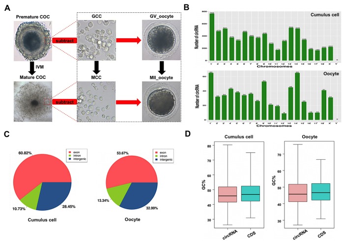
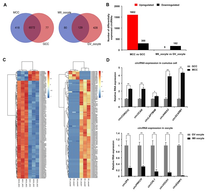
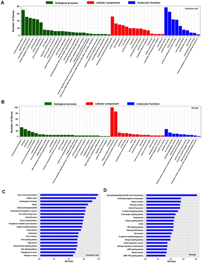
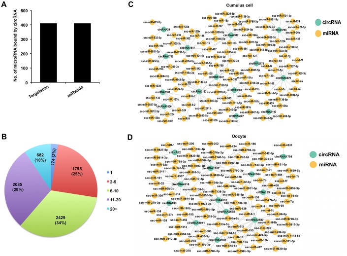
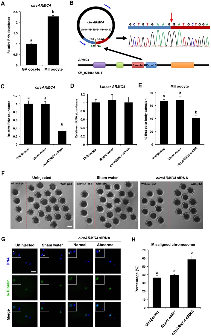
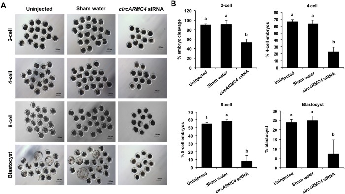
Thousands of circular RNAs (circRNAs) have been recently discovered in cumulus cells and oocytes from several species. However, the expression and function of circRNA during porcine oocyte meiotic maturation have been never examined. Here, we separately identified 7,067 and 637 circRNAs in both cumulus cells and oocytes via deep sequencing and bioinformatic analysis. Further analysis revealed that a faction of circRNAs is differentially expressed (DE) in a developmental stage-specific manner. The host genes of DE circRNAs are markedly enriched to multiple signaling pathways associated with cumulus cell function and oocyte maturation. Additionally, most DE circRNAs harbor several miRNA targets, suggesting that these DE circRNAs potentially act as miRNA sponge. Importantly, we found that maternal circARMC4 knockdown by siRNA microinjection caused a severely impaired chromosome alignment, and significantly inhibited first polar body extrusion and early embryo development. Taken together, these results demonstrate for the first time that circRNAs are abundantly and dynamically expressed in a developmental stage-specific manner in cumulus cells and oocytes, and maternally expressed circARMC4 is essential for porcine oocyte meiotic maturation and early embryo development.

Keywords: circular RNA; cumulus cells; meiotic maturation; oocytes; pig.


Conflict of interest statement

CONFLICTS OF INTEREST: The authors declare no conflicts of interest with regard to the study.

Figures

Figure 1
Figure 1
Characteristics of circRNAs expressed in porcine cumulus cells and oocytes. (A) Schematic illustration of experimental designs identifying circRNAs expressed in cumulus cells or oocytes before and after maturation. Premature COCs, cumulus cells before maturation (termed GCC), mature COCs, and cumulus cells after maturation (termed MCC) were collected respectively for RNA-seq. Of note, circRNAs expressed in the GCC were subtracted from these identical circRNAs expressed in the premature COCs to identify circRNAs expressed in GV oocytes, which are termed GV oocyte. Similarly, circRNAs expressed in MCC were subtracted from these identical circRNAs expressed in the mature COCs to identify circRNAs expressed in MII oocytes, which are termed MII oocyte. COCs, cumulus-oocyte complexes; GV, germinal vesicle; MII, metaphase II; IVM, in vitro maturation. Red dashed insets show cumulus cells before and after oocyte maturation at high magnification. (B) Chromosome distribution of circRNAs. Chromosome distribution of total circRNAs expressed in cumulus cells or oocytes was shown in the upper panel and bottom panel, respectively. (C) Genomic location of circRNAs. Genomic distribution of total circRNAs expressed in cumulus cells or oocytes was shown in the left panel and right panel, respectively. (D) GC enrichment of circRNAs and mRNAs. GC content of total circRNAs and mRNAs expressed in cumulus cells or oocytes was separately shown in the left panel and right panel.
Figure 2
Figure 2
Identification and validation of differentially expressed circRNAs (DECs) in both cumulus cells and oocytes during meiotic maturation. (A) Venn diagram of circRNAs identified in cumulus cells or oocytes. Cumulus cells and oocytes before and after meiotic maturation were pooled for RNA-seq. Expression levels of circRNAs in cumulus cells (left panel) and oocytes (right panel) were analyzed by means of a binominal statistical test. Overlapping circles present circRNAs that are common for cumulus cells or oocytes between two different stages. Non-overlapping circles indicate circRNAs that are specific for cumulus cells or oocytes before (pink) and after (blue) meiotic maturation. (B) The number of differentially expressed circRNAs in cumulus cells or oocytes before and after meiotic maturation. The results were considered statistically significant at Padjusted< 0.05 and log2 fold change ≥1. Red bars indicate up-regulated circRNAs; black bars denote down-regulated circRNAs. (C) Heatmap illustrating the expression patterns of differentially expressed circRNAs in cumulus cells (left panel) or oocytes (right panel) before and after meiotic maturation. The red blocks represent up-regulated circRNAs, and the blue blocks represent down-regulated circRNAs. The color scale of the heatmap indicates the expression level, where the brightest blue stands for -1.0 log2 fold change and the brightest red stands for 1.0 or 1.5 log2 fold change. (D) Validation of the selected differentially expressed circRNAs identified in both cumulus cells and oocytes. The several selected circRNAs were chosen from top up and top down-regulated circRNAs in cumulus cells or oocytes. Relative abundance of circRNAs in cumulus cells (upper panel) and oocytes (bottom panel) was determined by qPCR. The data were normalized against endogenous housekeeping gene EF1α1, and the value for cumulus cells or oocytes at GV stage was set as one. The data are shown as mean ± S.E.M. Statistical analysis was performed using t-student test. Values with asterisks vary significantly, *P < 0.05, **P < 0.01.
Figure 3
Figure 3
GO and KEGG analysis of host genes of differentially expressed circRNAs (DECs) in both cumulus cells and oocytes during meiotic maturation. (A) GO analysis of the top enriched terms of the differentially expressed circRNA hosting genes identified in cumulus cells. Host genes of differentially expressed circRNAs were classified into three categories of the GO classification (blue bars: biological processes, green bars: cellular components and orange bars: molecular functions). (B) KEGG analysis of the top enriched signaling pathways of the differentially expressed circRNA hosting genes identified in cumulus cells. (C) GO analysis of the top enriched terms of the differentially expressed circRNA hosting genes identified in oocytes. Host genes of differentially expressed circRNAs were classified into three categories of the GO classification (blue bars: biological processes, green bars: cellular components and orange bars: molecular functions). (D) KEGG analysis of the top enriched signaling pathways of the differentially expressed circRNA hosting genes identified inoocytes.
Figure 4
Figure 4
Analysis of interaction between DECs and miRNAs in both cumulus cells and oocytes. (A) Analysis of number of miRNAs for circRNAs by Targetscan and miRanda. (B) Analysis of the proportion of circRNA processing different numbers of miRNA targets. (C) Analysis for predicted targeted miRNAs of the selected DECs identified in cumulus cells. The selected circRNAs were chosen from top DECs in cumulus cells. Blue circles represent circRNA, and yellow circles represent miRNA. (D) Analysis for predicted targeted miRNAs of the selected DECs identified in oocytes. The selected circRNAs were chosen from top DECs in oocytes. Blue circles represent circRNA, and yellow circles represent miRNA.
Figure 5
Figure 5
Effect of circARMC4 knockdown on oocyte meiotic maturation and chromosome alignment. (A) CircARMC4 expression in both GV oocytes and MII oocytes. Relative expression of circARMC4 was determined by qPCR. The data are analyzed using student’s t test and are shown as mean ± S.E.M. Different letters on the bars indicate significant differences (P < 0.05). (B) Schematic illustration showed the ARMC4 exon 14 and exon 15 circularization forming circARMC4 (blue arrow). The presence of circARMC4 was validated by qPCR, followed by Sanger sequencing. Red arrow represents “head-to-tail” circARMC4 splicing sites. The expression levels of circARMC4 (C) and linear ARMC4 (D) in the MII oocytes derived from GV oocytes. GV oocytes were injected with circARMC4 siRNA, followed by maturation in vitro for 44 h. Oocytes injected with water and uninjected oocytes were served as a sham control and a blank control, respectively. One hundred matured oocytes were collected for qPCR analysis. Relative abundance of circARMC4 and linear ARMC4 was determined by qPCR from four independent replicates. The data were normalized against endogenous housekeeping gene EF1α1 and the value for the blank control was set as one. The data are shown as mean ± S.E.M. One-way ANOVA was used to analyze the data and different letters on the bars indicate significant differences (P < 0.05). (E) Analysis of the rate of oocyte maturation. The number of oocytes with first polar body after in vitro maturation for 44 h was recorded and the rate of first polar body extrusion was statistically analyzed by one-way ANOVA. The experiment was repeated four times with at least 100 oocytes per group. The data are shown as mean ± S.E.M and different letters on the bars indicate significant differences (P < 0.05). (F) Representative images of oocytes after in vitro maturation. The oocytes without pb1 and the oocytes with pb1 were shown in both the left side and the right side of each image, respectively. Scale bar: 100 μm. (G) Spindle and chromosome morphology in MII oocytes. Matured oocytes were stained for α-tubulin (green) and DAPI (blue). Shown are representative images obtained using confocal laser-scanning microscopy. The experiment was independently repeated three times with at least 40 oocytes per group. Bottom panel in each group shows the merged images between α-tubulin and DNA. White square insets indicate both spindles and chromosomes at a more focused view. Asterisks indicate chromosomes. Scale bar: 50 μm. (H) Analysis of the percentage of oocytes with abnormal chromosome morphology. The chromosome morphology of MII oocytes was scored according to a published method. The data were statistically analyzed by one-way ANOVA. The data are shown as mean ± S.E.M and different superscripts on the bars indicate significant differences (P < 0.05).
Figure 6
Figure 6
Effect of circARMC4 knockdown on developmental competence of porcine early embryos. (A) Representative brightfield images of embryos at different developmental stages. GV oocytes were injected with siRNA or water and GV oocytes without any treatment, followed by maturation in vitro for 44 h. Oocytes with first polar body were parthenogenetically activated (n=51, 57, 28) and cultured up to the blastocyst stage. The brightfield images of 2-cell, 4-cell, 8-cell embryo and blastocyst were captured by epifluorescence microscopy. Scale bar: 100 μm. (B) Analysis of the developmental rate of embryos at different developmental stages. The number of embryos at different developmental stages was recorded (2-cell: n=46, 52, 14; 4-cell: n=34, 38, 6; 8-cell: n=28, 33, 2; blastocyst: n=12, 14, 2) and the corresponding data were statistically analyzed by one-way ANOVA. All experiment was repeated three times. The data are shown as mean ± S.E.M and different letters on the bars indicate significant differences (P < 0.05).

Similar articles

Cited by

References

    1. Conti M, Franciosi F. Acquisition of oocyte competence to develop as an embryo: integrated nuclear and cytoplasmic events. Hum Reprod Update. 2018; 24:245–66. 10.1093/humupd/dmx040 - DOI - PMC - PubMed
    1. Russell DL, Gilchrist RB, Brown HM, Thompson JG. Bidirectional communication between cumulus cells and the oocyte: old hands and new players? Theriogenology. 2016; 86:62–68. 10.1016/j.theriogenology.2016.04.019 - DOI - PubMed
    1. Gilchrist RB, Ritter LJ, Armstrong DT. Oocyte-somatic cell interactions during follicle development in mammals. Anim Reprod Sci. 2004; 82-83:431–46. 10.1016/j.anireprosci.2004.05.017 - DOI - PubMed
    1. Gilchrist RB, Lane M, Thompson JG. Oocyte-secreted factors: regulators of cumulus cell function and oocyte quality. Hum Reprod Update. 2008; 14:159–77. 10.1093/humupd/dmm040 - DOI - PubMed
    1. Nagai T. The improvement of in vitro maturation systems for bovine and porcine oocytes. Theriogenology. 2001; 55:1291–301. 10.1016/S0093-691X(01)00483-6 - DOI - PubMed

Publication types

LinkOut - more resources