

Smoking Attenuates Efficacy of Penehyclidine Hydrochloride in Acute Respiratory Distress Syndrome Induced by Lipopolysaccharide in Rats
- PMID: 31562811
- PMCID: PMC6784682
- DOI: 10.12659/MSM.917037
Smoking Attenuates Efficacy of Penehyclidine Hydrochloride in Acute Respiratory Distress Syndrome Induced by Lipopolysaccharide in Rats
Abstract
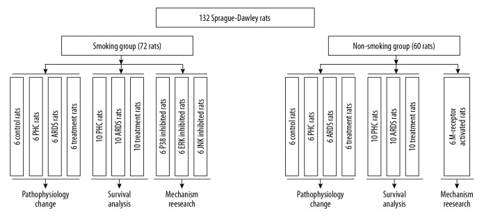
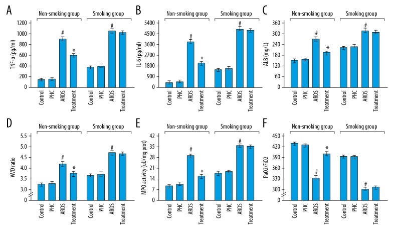
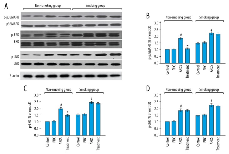
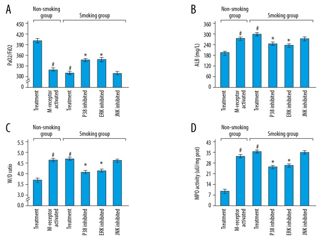
BACKGROUND Penehyclidine hydrochloride is a novel drug for acute respiratory distress syndrome. The aim of the study was to reveal the impact of smoking on the efficacy of the drug in rats with acute respiratory distress syndrome. MATERIAL AND METHODS A 132 Sprague-Dawley rats were used in this study; 72 rats were used in the smoking models. Penehyclidine hydrochloride (3 mg/kg) was injected to induce acute respiratory distress syndrome. Rats were divided into the smoking group and the non-smoking group; these 2 groups were subdivided according to different treatments. The arterial blood gas analysis (PaO₂/FiO₂) and extent of pneumonedema (wet-to-dry weight ratio) was analyzed to evaluate disease severity. Expressions of mitogen-activated protein kinases (p-p38MAPK, p38MAPK, p-ERK, ERK, p-JNK, and JNK) in lung tissue were measured using western blot assay. RESULTS Penehyclidine hydrochloride improved the pneumonedema (wet-to-dry weight ratio) and hyoxemia (PaO₂/FiO₂) of the disease in non-smoking group (P<0.001, P<0.001 respectively), but not in smoking group (P=0.244, P=0.424 respectively). The drug inhibited the expressions of phospho-p38MAPK and phospho-ERK in non-smoking group (P<0.001, P<0.001 respectively), but not in smoking group (P=0.350, P=0.507 respectively). In the smoking group, blocking the phospho-p38MAPK or phospho-ERK signal pathway by their inhibitors showed a better therapeutic effect on the pneumonedema and hyoxemia compared with the use of penehyclidine hydrochloride (phospho-p38MAPK: P=0.004, P=0.010 respectively; phospho-ERK: P=0.022, P=0.004 respectively). CONCLUSIONS The study confirmed the protective effect of penehyclidine hydrochloride in acute respiratory distress syndrome, mainly in the non-smoking group, which might be explained by the fact that phospho-p38MAPK and phospho-ERK signal pathways were difficult to inhibit by the drug in the smoking group.
Conflict of interest statement
None.
Figures

References
-
- ARDS Definition Task Force. Ranieri VM, Rubenfeld GD, Thompson BT, et al. Acute respiratory distress syndrome: The Berlin Definition. JAMA. 2012;307:2526–33. - PubMed
-
- Ferguson ND, Fan E, Camporota L, et al. The Berlin definition of ARDS: An expanded rationale, justification, and supplementary material. Intensive Care Med. 2012;38:1573–82. - PubMed
-
- Fan E, Brodie D, Slutsky AS. Acute respiratory distress syndrome: Aadvances in diagnosis and treatment. JAMA. 2018;319:698–710. - PubMed
-
- Aeffner F, Bolon B, Davis IC. Mouse models of acute respiratory distress syndrome: A review of analytical approaches, pathologic features, and common measurements. Toxicol Pathol. 2015;43:1074–92. - PubMed
MeSH terms
Substances
LinkOut - more resources
Full Text Sources
Research Materials
Miscellaneous
