. 2019 Dec 15:317:1-12.
doi: 10.1016/j.toxlet.2019.09.013. Epub 2019 Sep 25.

Acrylonitrile butadiene styrene (ABS) and polycarbonate (PC) filaments three-dimensional (3-D) printer emissions-induced cell toxicity

Affiliations

Acrylonitrile butadiene styrene (ABS) and polycarbonate (PC) filaments three-dimensional (3-D) printer emissions-induced cell toxicity

Mariana T Farcas et al. Toxicol Lett. .

Abstract

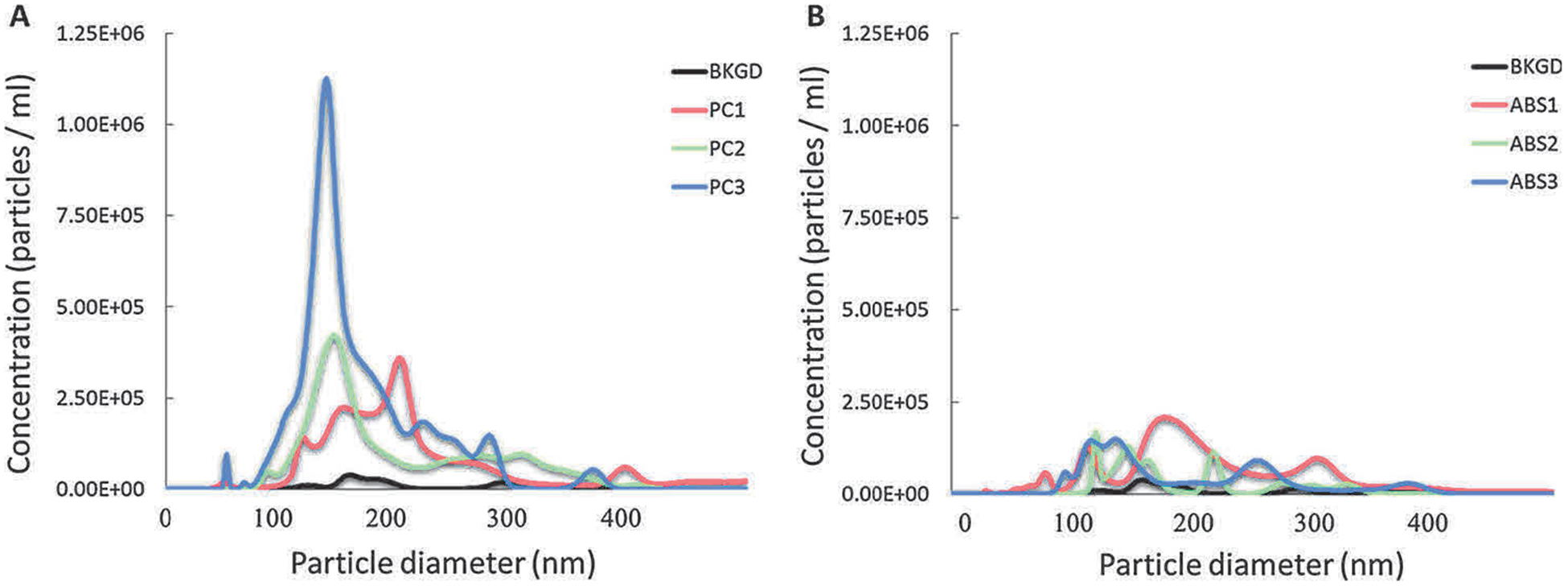
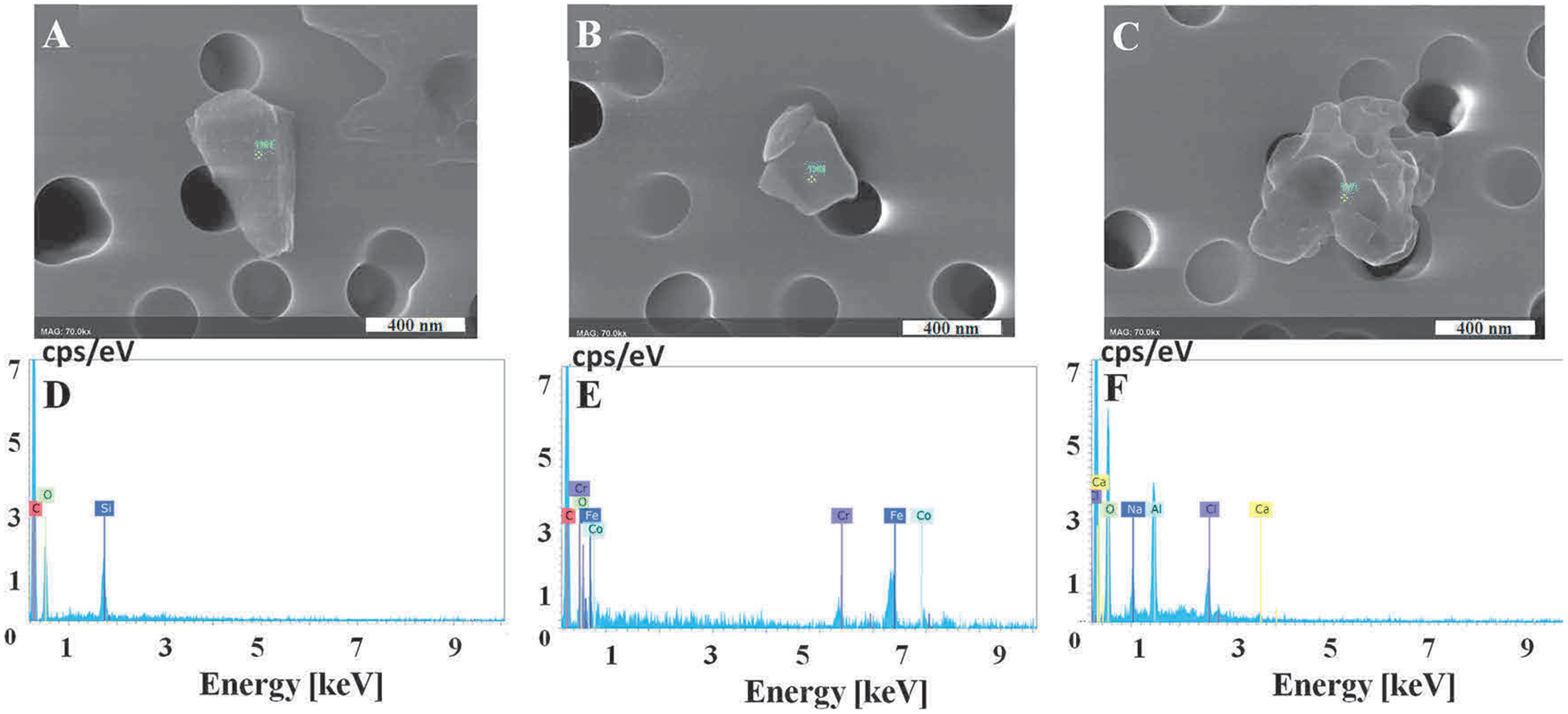
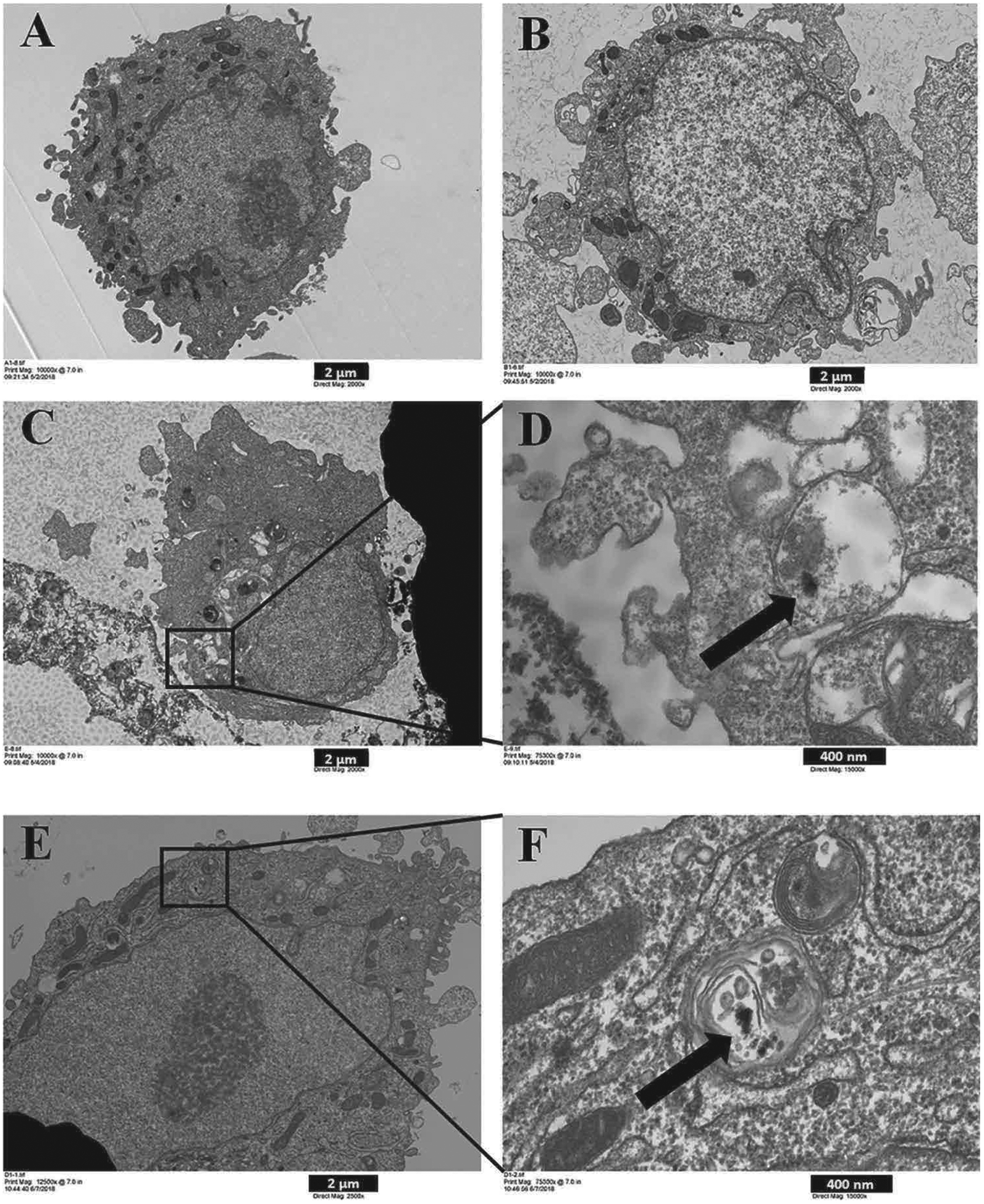
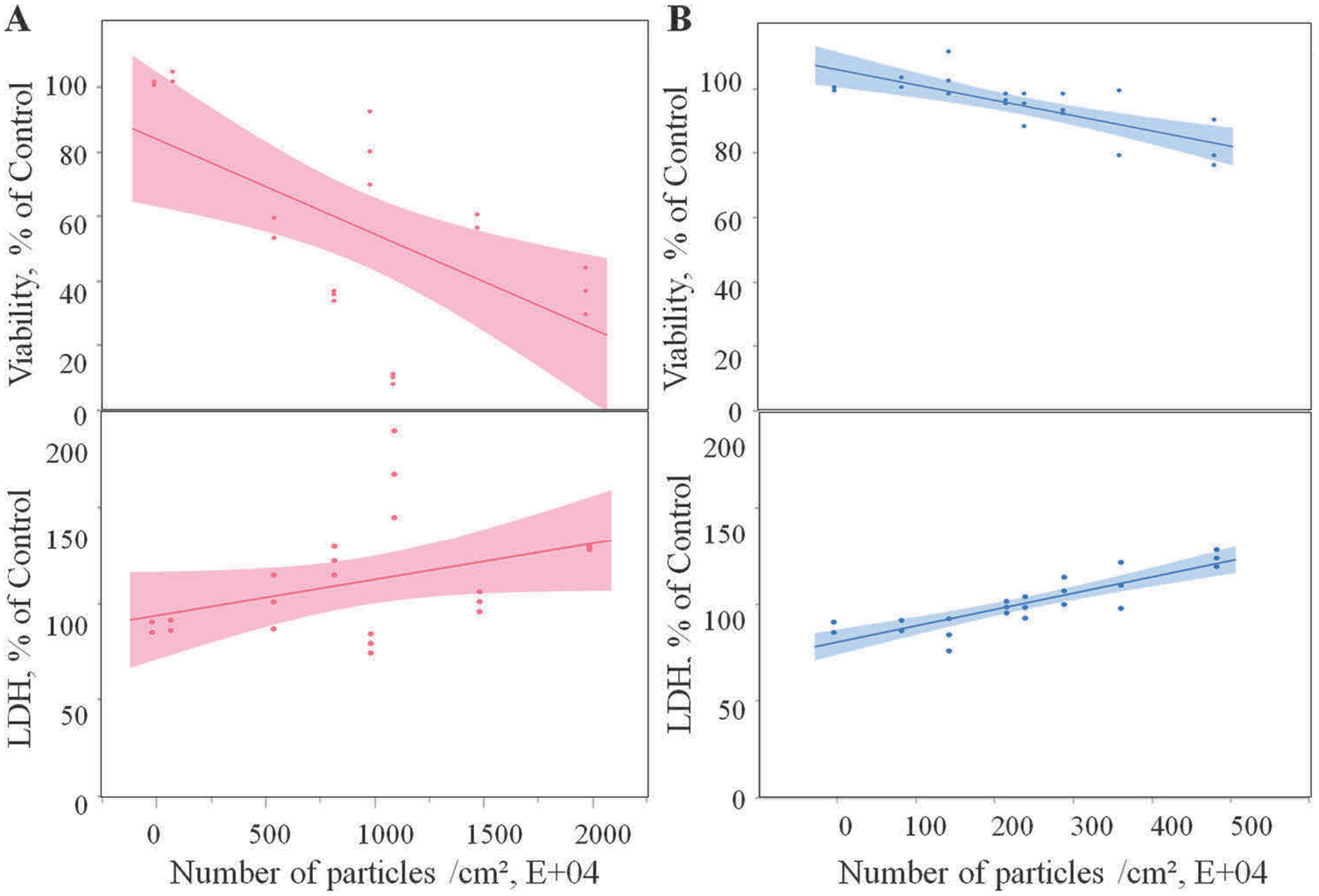
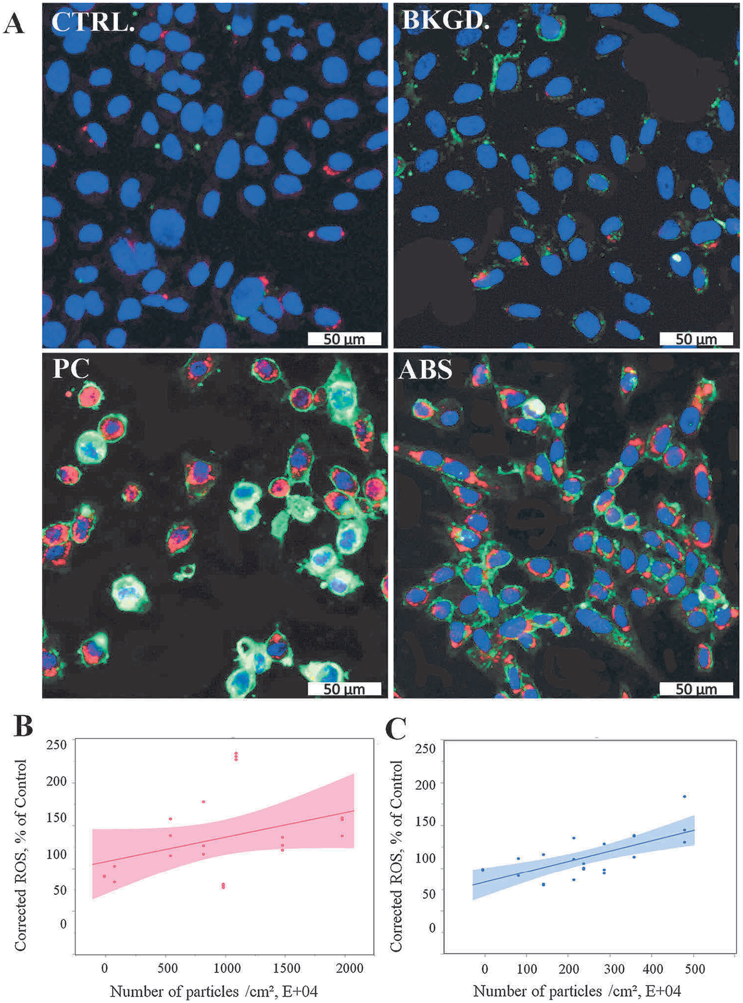
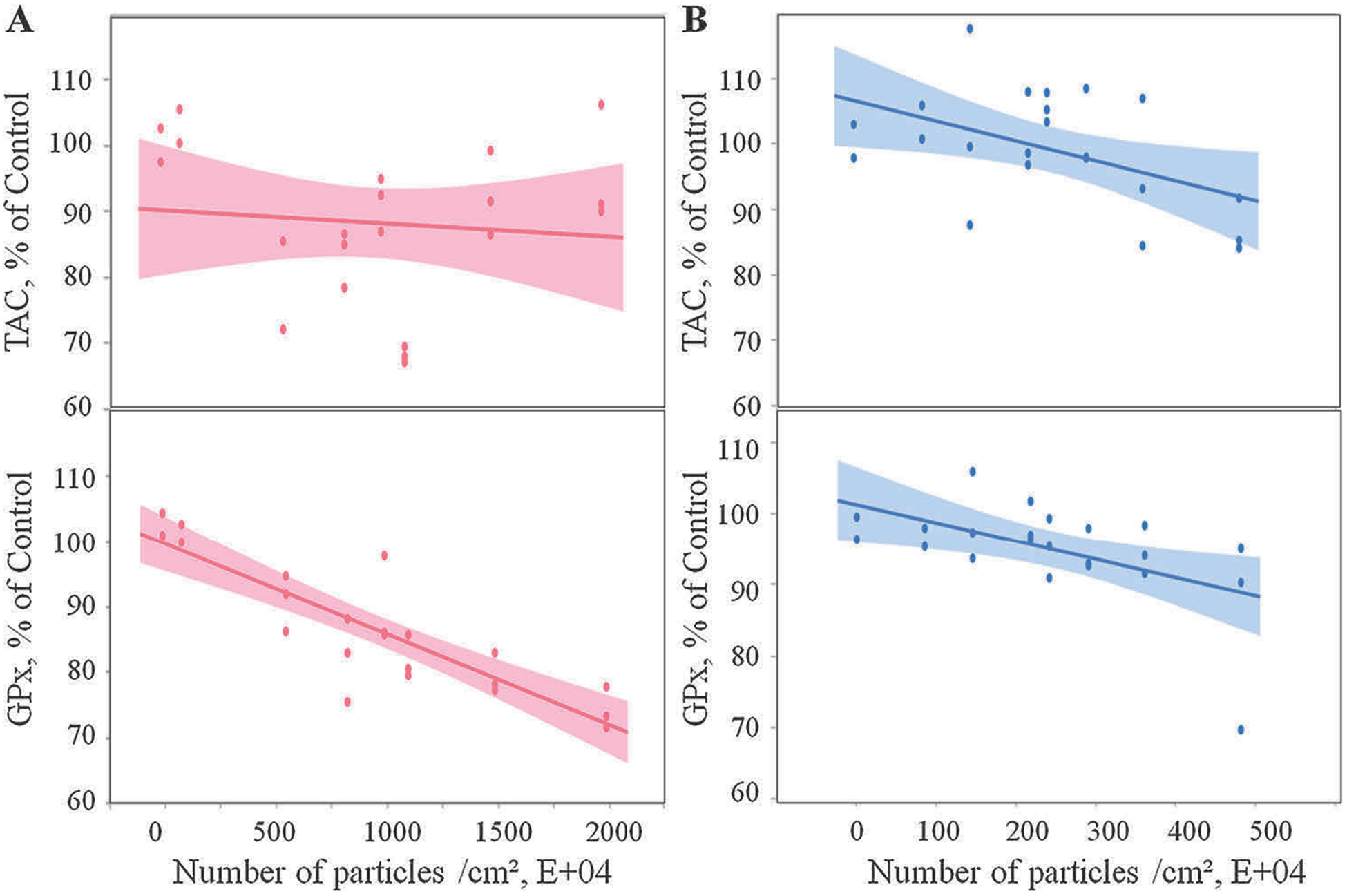
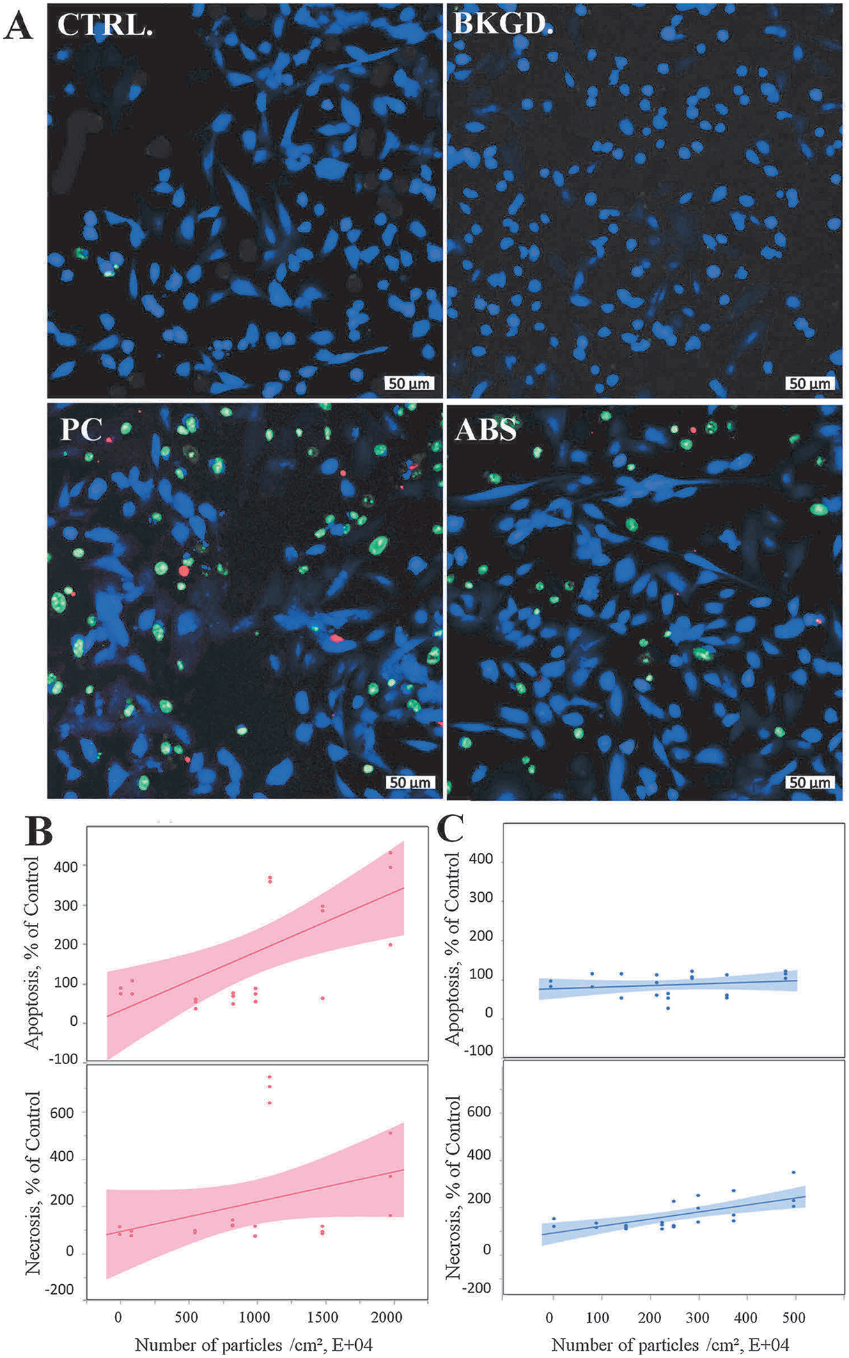
During extrusion of some polymers, fused filament fabrication (FFF) 3-D printers emit billions of particles per minute and numerous organic compounds. The scope of this study was to evaluate FFF 3-D printer emission-induced toxicity in human small airway epithelial cells (SAEC). Emissions were generated from a commercially available 3-D printer inside a chamber, while operating for 1.5 h with acrylonitrile butadiene styrene (ABS) or polycarbonate (PC) filaments, and collected in cell culture medium. Characterization of the culture medium revealed that repeat print runs with an identical filament yield various amounts of particles and organic compounds. Mean particle sizes in cell culture medium were 201 ± 18 nm and 202 ± 8 nm for PC and ABS, respectively. At 24 h post-exposure, both PC and ABS emissions induced a dose dependent significant cytotoxicity, oxidative stress, apoptosis, necrosis, and production of pro-inflammatory cytokines and chemokines in SAEC. Though the emissions may not completely represent all possible exposure scenarios, this study indicate that the FFF could induce toxicological effects. Further studies are needed to quantify the detected chemicals in the emissions and their corresponding toxicological effects.

Keywords: Emerging technologies; In vitro toxicity; Inflammatory response; Printer emitted nanoparticles.


Conflict of interest statement

Declaration of Competing Interest

The authors declare no competing financial interest.

Figures

Fig. 1.
Fig. 1.
Number-based size distribution of 3-D printer-emitted particles collected in the cell culture medium. 3-D printer emissions using PC (A) and ABS (B) were collected in cell culture medium, followed by analysis using NTA.
Fig. 2.
Fig. 2.
Representative images of 3-D printer-emitted particles collected in the cell culture medium indicating surface morphology and elemental composition. 3-D printer emissions were collected in cell culture medium, passed through a track-etched polycarbonate filter, and analyzed using FE-SEM (A–C) and EDX (D–F). (A) Background, (B) PC, (C) ABS. D–F are representative elemental analysis spectra for A–C. Magnification at 70k × for all images, scale bar at 400 nm.
Fig. 3.
Fig. 3.
Uptake by SAEC of 3-D printer-emitted particles collected in the cell culture medium. SAEC were treated with medium containing 3-D printer emissions for 24 h, followed by TEM image analysis. (A) Control, (B) Background, (C) 100% PC, (D) C inset, (E) 100% ABS, and (F) E inset. A, B, and C images are at 2,000x magnification, and image E at 2,500x magnification. D and F images are at 15,000x magnification. A, B, C, and E scale bar at 2 μm; D, F scale bar at 400 nm.
Fig. 4.
Fig. 4.
Cytotoxicity of the 3-D printer-emitted particles collected in the cell culture medium: cell viability and LDH activity following exposure to PC (A) and ABS (B) emissions. Experiments were performed in three independent experiments with n = 6 replicates each. The regressions lines illustrate a significantly (p < 0.0001) decreased dose-response relationship between cell proliferation and the numbers of the PC and ABS 3-D printer-emitted particles, which correlated with a significant (p < 0.0001) dose-dependent increase in the LDH activity in the supernatants. The shaded area represents the 95% confidence interval around the regression line.
Fig. 5.
Fig. 5.
ROS production following exposure to 3-D printer-emitted particles collected in the cell culture medium. SAEC were stained with a cocktail consisting of 5 μM CellROX™ Deep Red reagent, 5 μg/mL of CellMask™ Orange plasma membrane stain, and 1 μM Hoechst 33342 fluorescent nuclear stain. (A) Representative confocal images of untreated cells, and cells exposed to background, PC, and ABS collected emissions for 24 h. Quantitative ROS levels of PC (B) and ABS (C) using high content screening. Magnification at 20× and scale bars are at 50 μm for all images. Experiments were performed in three independent experiments with n = 6 replicates each. The regressions lines illustrate a significantly (p < 0.0001) increased dose-response relationship between ROS production and the numbers of the PC and ABS 3-D printer-emitted particles. The shaded area represents the 95% confidence interval around the regression line.
Fig. 6.
Fig. 6.
Oxidative stress response following 3-D printer-emitted particles collected in the cell culture medium: total antioxidant capacity and glutathione peroxidase activity following exposure to PC (A) and ABS (B) emissions. Experiments were performed in three independent experiments with n = 3 replicates each. The regressions lines illustrate a significantly (p < 0.0001) decreased dose-response relationship between total antioxidant capacity and glutathione peroxidase activity and the numbers of the PC and ABS 3-D printer-emitted particles. The shaded area represents the 95% confidence interval around the regression line.
Fig. 7.
Fig. 7.
3-D printer-emitted particles collected in the cell culture medium increase apoptosis and necrosis. SAEC were stained with a cocktail consisting of 2 μL of Apopxin™ Red apoptosis stain, 1 μL of 200X Nuclear Green™ DCS1 necrotic stain, and 1 μL of CytoCalcein™ Violet 450 cytoplasmic stain. (A) Representative confocal images of untreated cells, and cells exposed to background, PC, and ABS collected emissions for 24 h. Quantitative apoptosis and necrosis analysis of PC (B) and ABS (C) using high content screening image analysis. Magnification at 10× and scale bars are at 50 μm for all images. Experiments were performed in three independent experiments with n = 6 replicates each. The regressions lines illustrate a significantly (p < 0.0001) increased dose-response relationship between apoptotic or necrotic events and the numbers of the PC and ABS 3-D printer-emitted particles. The shaded area represents the 95% confidence interval around the regression line.

References

    1. ASTM-D6670, 2013. ASTM D6670: Standard Practice for Full-scale Chamber Determination of Volatile Organic Emissions from Indoor Materials/Products. ASTM International, West Conshohocken, PA.
    1. Azimi P, Zhao D, Pouzet C, Crain NE, Stephens B, 2016. Emissions of ultrafine particles and volatile organic compounds from commercially available desktop three-dimensional printers with multiple filaments. Environ. Sci. Technol 50, 1260–1268. - PubMed
    1. Barnes PJ, 2008. The cytokine network in asthma and chronic obstructive pulmonary disease. J. Clin. Invest 118, 3546–3556. - PMC - PubMed
    1. Barnes PJ, 2009. The cytokine network in chronic obstructive pulmonary disease. Am. J. Respir. Cell Mol. Biol 41, 631–638. - PubMed
    1. Berda-Haddad Y, Robert S, Salers P, Zekraoui L, Farnarier C, Dinarello CA, Dignat-George F, Kaplanski G, 2011. Sterile inflammation of endothelial cell-derived apoptotic bodies is mediated by interleukin-1alpha. Proc. Natl. Acad. Sci. U. S. A 108, 20684–20689. - PMC - PubMed

MeSH terms

LinkOut - more resources