. 2020 Apr;77(4):420-433.
doi: 10.1016/j.eururo.2019.09.006. Epub 2019 Sep 26.

A Consensus Molecular Classification of Muscle-invasive Bladder Cancer

Collaborators, Affiliations

A Consensus Molecular Classification of Muscle-invasive Bladder Cancer

Aurélie Kamoun et al. Eur Urol. 2020 Apr.

Abstract

Background: Muscle-invasive bladder cancer (MIBC) is a molecularly diverse disease with heterogeneous clinical outcomes. Several molecular classifications have been proposed, but the diversity of their subtype sets impedes their clinical application.

Objective: To achieve an international consensus on MIBC molecular subtypes that reconciles the published classification schemes.

Design, setting, and participants: We used 1750 MIBC transcriptomic profiles from 16 published datasets and two additional cohorts.

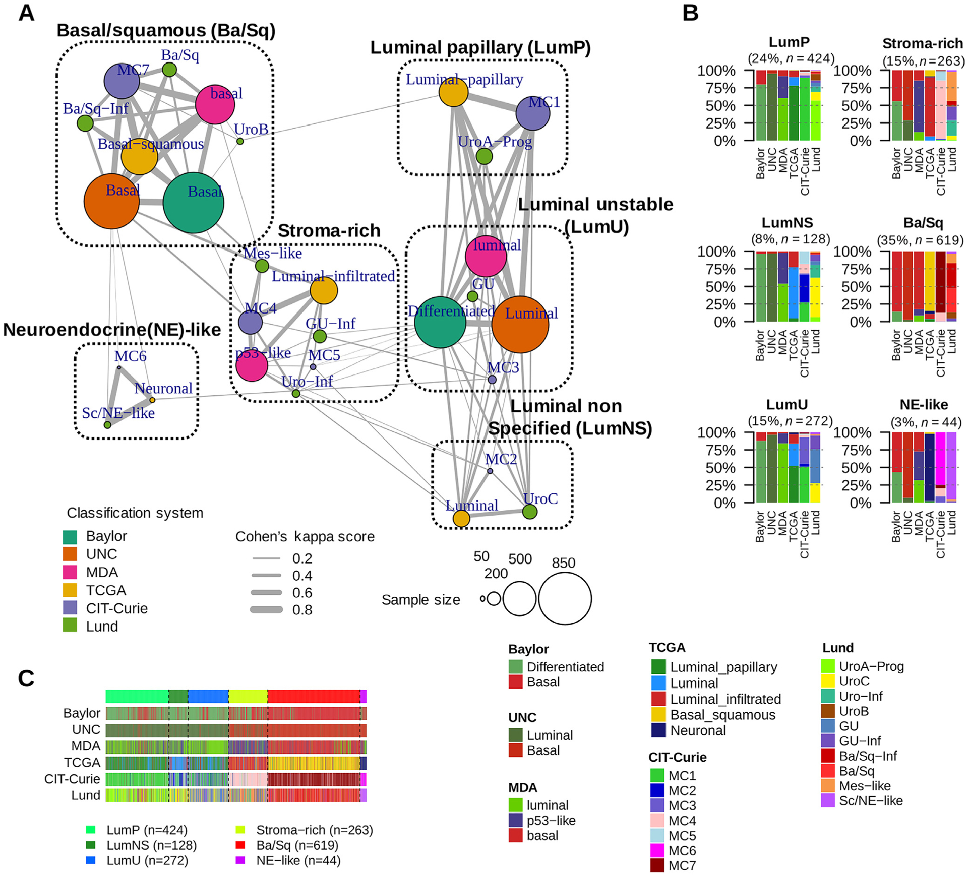
Outcome measurements and statistical analysis: We performed a network-based analysis of six independent MIBC classification systems to identify a consensus set of molecular classes. Association with survival was assessed using multivariable Cox models.

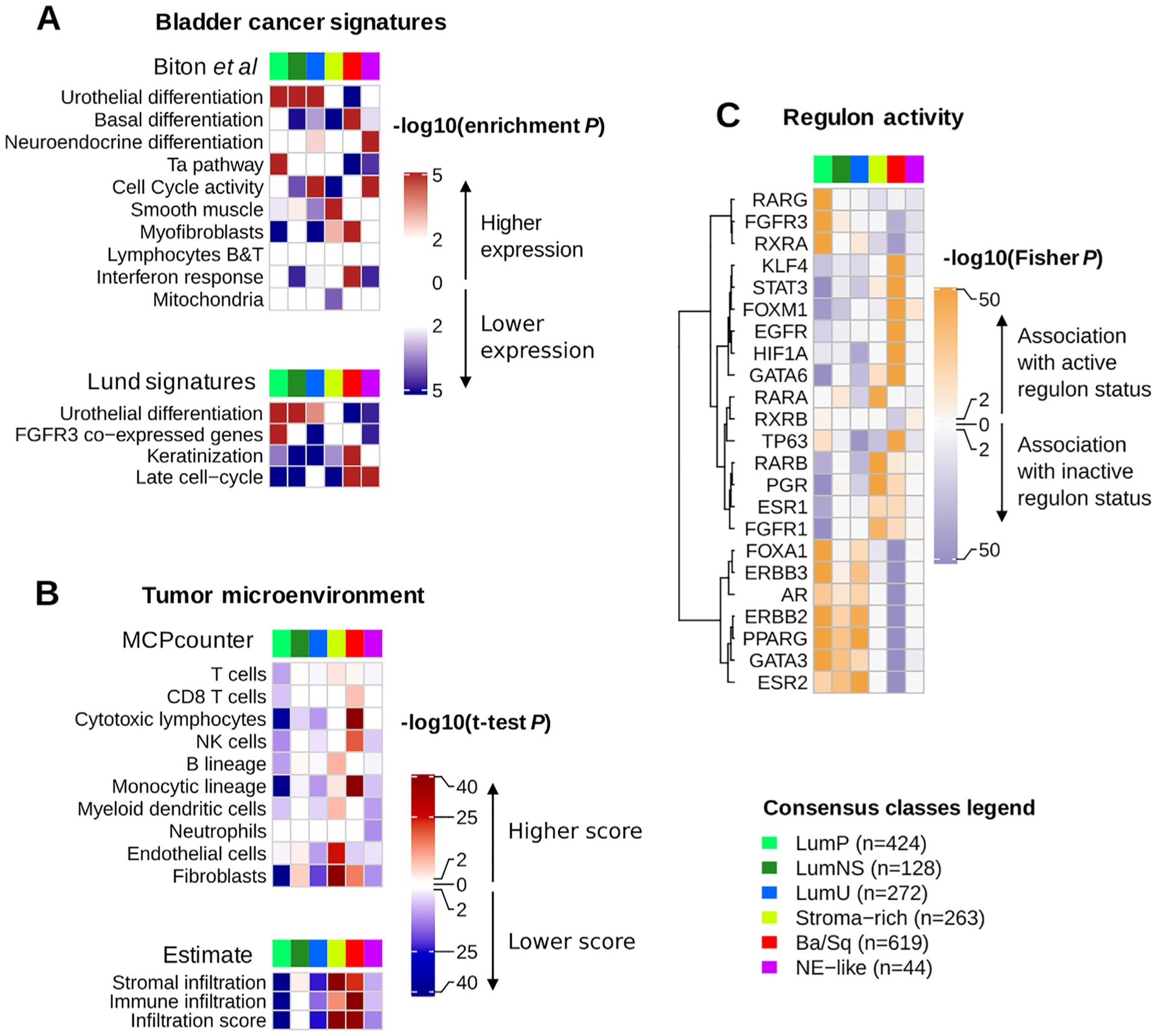
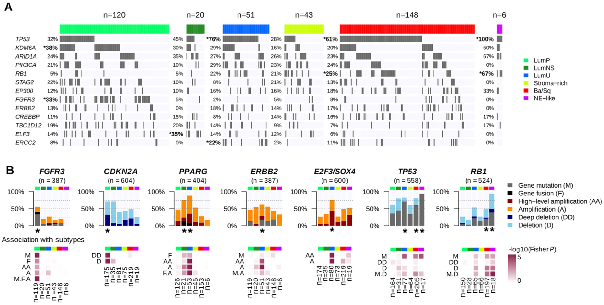
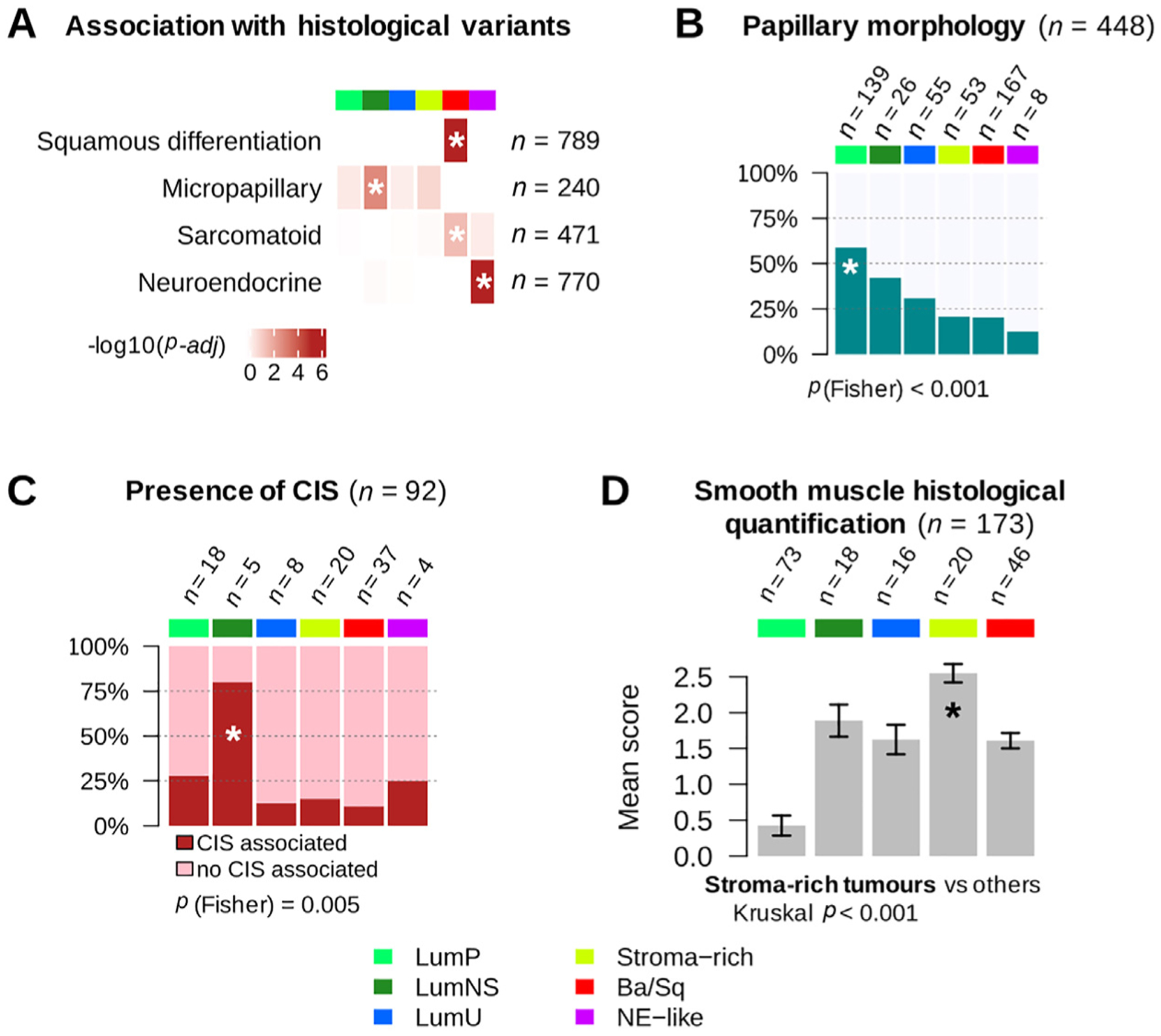
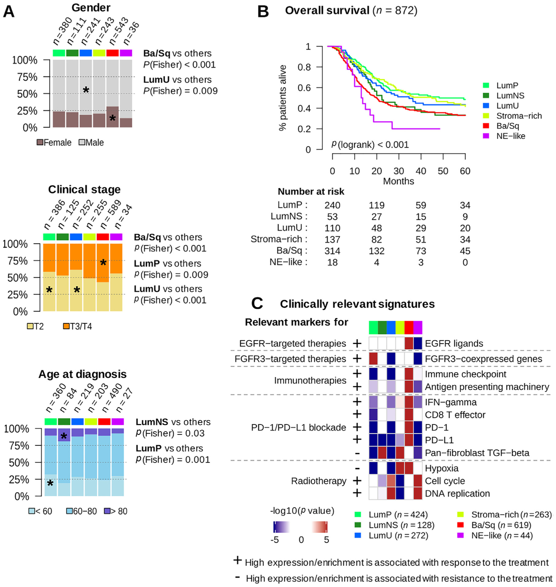
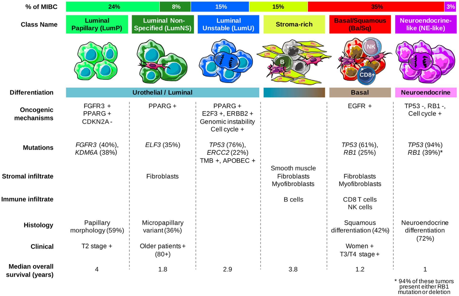
Results and limitations: We report the results of an international effort to reach a consensus on MIBC molecular subtypes. We identified a consensus set of six molecular classes: luminal papillary (24%), luminal nonspecified (8%), luminal unstable (15%), stroma-rich (15%), basal/squamous (35%), and neuroendocrine-like (3%). These consensus classes differ regarding underlying oncogenic mechanisms, infiltration by immune and stromal cells, and histological and clinical characteristics, including outcomes. We provide a single-sample classifier that assigns a consensus class label to a tumor sample's transcriptome. Limitations of the work are retrospective clinical data collection and a lack of complete information regarding patient treatment.

Conclusions: This consensus system offers a robust framework that will enable testing and validation of predictive biomarkers in future prospective clinical trials.

Patient summary: Bladder cancers are heterogeneous at the molecular level, and scientists have proposed several classifications into sets of molecular classes. While these classifications may be useful to stratify patients for prognosis or response to treatment, a consensus classification would facilitate the clinical use of molecular classes. Conducted by multidisciplinary expert teams in the field, this study proposes such a consensus and provides a tool for applying the consensus classification in the clinical setting.

Keywords: Consensus; Molecular taxonomy; Muscle-invasive bladder cancer; Transcriptomic classifier.


Figures

Fig. 1 –
Fig. 1 –
The six consensus classes and their relationships to input molecular subtypes. (A) MCL-clustered network. The six-consensus class solution obtained with MCL clustering on the Cohen’s kappa-weighted network is represented by the six cliques surrounded by black dotted rectangles (see the Supplementary material [Note] for the naming of consensus classes). The circles inside each clique symbolize the input subtypes associated with each consensus class and are colored according to their matching classification system. Circle size is proportional to the number of samples assigned to the subtype. Edge width between subtypes is proportional to the Cohen’s kappa score, which assesses the level of agreement between two classification schemes. (B) Input subtypes repartitioned among each consensus class. Consensus classes were predicted on 1750 MIBC samples using the single-sample classifier described in the Supplementary material (Methods). Here, the samples are grouped by their predicted consensus class labels: LumP, LumNS, LumU, stroma-rich, Ba/Sq, and neuroendocrine (NE)-like. For each consensus class, a bar plot shows the proportion of samples assigned in each input subtype of each input classification system. See also Supplementary Fig. 2 for additional visualization of consensus class distributions across input subtypes and across datasets. (C) Relationship between subtyping results from the six input classification schemes. Samples are ordered by predicted consensus classes. Ba/Sq = basal/squamous; LumNS = luminal nonspecified; LumP = luminal papillary; LumU = luminal unstable; MCL = Markov cluster algorithm; MDA = MD Anderson Cancer Center; MIBC = muscle-invasive bladder cancer; TCGA = the Cancer Genome Atlas; UNC = University of North Carolina.
Fig. 2 –
Fig. 2 –
Characterization of tumor and stroma signals using published mRNA signatures and regulon analysis. Descriptions of gene sets and detailed statistics are available in Supplementary Table 3. (A) We performed a gene set analysis (GSA; see the Supplementary material, Methods) in each dataset to test the significance of differential expression of specific bladder cancer-related signatures in each consensus class compared with the others. The heatmaps show Stouffer combined GSA p values over all datasets. The upper panel refers to bladder cancer gene sets extracted from the ICA components described in the study by Biton et al. [25] (see the Supplementary material, Methods). The lower panel displays other bladder cancer-specific signatures retrieved from the literature: urothelial differentiation, keratinization, and late cell-cycle signatures from the study of Eriksson et al. [24], and an FGFR3 coexpressed signature from the study of Sjödahl et al. [4]. (B) We used two mRNA-based computational tools to characterize tumor microenvironments : ESTIMATE (R package, v1.1.0) infers the presence of stromal cells (stromal infiltration) and the infiltration of immune cells (immune infiltration) in a tumor sample using two curated gene signatures described by Yoshihara et al. [26]; MCPcounter (R package, v1.0.13) uses biologically validated transcriptomic markers of specific immune and stromal cell subpopulations to quantify the presence of these populations in a tumor sample [27]. We ran MCPcounter and ESTIMATE independently on each dataset, and used t tests to compare scores for each consensus class relative to the others. The heatmaps show Stouffer combined t test p values over all datasets. (C) We computed discrete regulon status (1 for active regulon status, 0 for undefined status, and −1 for inactive regulon status) in each dataset, as described in the Supplementary material (Methods) and in the work of Robertson et al. [9]. We evaluated the association between each regulon status and each consensus class using Fisher exact tests; the heatmap illustrates the resulting p values. Ba/Sq = basal/squamous; ICA = independent component analysis; LumNS = luminal nonspecified; LumP = luminal papillary; LumU = luminal unstable; NE = neuroendocrine; NK = natural killer.
Fig. 3 –
Fig. 3 –
Genomic alterations associated with consensus classes. (A) We used the available exome data from 388 TCGA MIBC samples to study the association between consensus classes and specific gene mutations (see Supplementary Table 4 and Supplementary Fig. 3). The panel displays the 13 genes with MutSig q values <0.02 found in >10% of all tumors. Gene mutations that were significantly enriched in one consensus class are marked by an asterisk. (B) Combined genomic alterations associated with seven bladder cancer-associated genes and statistical association with consensus classes. Upper panels: main alteration types after aggregating CNA profiles (see Supplementary Table 5) from CIT (n = 87), Iyer (n = 58), Sjödahl (n = 29), Stransky (n = 22), and TCGA (n = 404) data; exome profiles (n = 388) and FGFR3 and PPARG fusion data (n = 404) from TCGA data; CDKN2A and RB1 MLPA data from CIT (n = 86 and n = 85, respectively) and Stransky (n = 16 and n = 13, respectively) data; FGFR3 mutation data from MDA (n = 66), CIT (n = 87), Iyer (n = 39), Sjödahl (n = 28), and Stransky (n = 35); TP53 mutation data from MDA (n = 66), CIT (n = 87), Iyer (n = 39), Sjödahl (n = 28), and Stransky (n = 19); and RB1 mutation data from MDA (n = 66), CIT (n = 85), Iyer (n = 39), and Stransky (n = 13). Lower panels: associations between each consensus class, each type of gene alteration, and the combined alterations were evaluated by Fisher’s exact test. Consensus classes significantly enriched with alterations of these candidate genes are marked with a black asterisk. Ba/Sq = basal/squamous; CIT = Cartes d’Identité des Tumeurs; CNA = copy number aberration; LumNS = luminal nonspecified; LumP = luminal papillary; LumU = luminal unstable; MDA = MD Anderson Cancer Center; MIBC = muscle-invasive bladder cancer; NE = neuroendocrine; TCGA = the Cancer Genome Atlas.
Fig. 4 –
Fig. 4 –
Histopathological associations with consensus classes. (A) Histological variant over-representation within each consensus class. One-sided Fisher exact tests were performed for each class and histological pattern; asterisks indicate a significant association between a consensus class and a histological feature (p < 0.05). Pathological review of histological variants was available for several cohorts: squamous differentiation was evaluated in CIT (n = 75), MDA (n = 46), Sjödahl2012 (n = 23), Sjödahl2017 (n = 239), and TCGA (n = 406) cohorts; neuroendocrine variants were reviewed in CIT (n = 75), MDA (n = 46), Sjödahl2017 (n = 243), and TCGA (n = 406) cohorts; micropapillary variants were reviewed in CIT (n = 75), MDA (n = 46), and TCGA cohorts (n = 118 FFPE tumor slides from TCGA were reviewed by Y.A. and J.F. for this study). Results are displayed on the heatmap as −log10(adj Fisher’s p). Detailed sample counts within each class are given in Supplementary Fig. 4. (B) Occurrence of papillary morphology in tumors from the TCGA cohort (n = 401) and the CIT cohort (n = 47). (C) Proportion of samples with associated CIS within each consensus class in tumors from the CIT cohort (n = 84) and the Dyrskjøt cohort (n = 8). (D) Smooth muscle infiltration from images for 173 tumor slides from the TCGA cohort. Each sample was assigned a semiquantitative score ranging from 0 to 3 (0 = absent, 1 = low, 2 = moderate, and 3 = high) to quantify the presence of large smooth muscle bundles. The bar plot shows means and standard errors for each class. Ba/Sq = basal/squamous; CIS = carcinoma in situ; CIT = Cartes d’Identité des Tumeurs; FFPE = formalin-fixed paraffin-embedded; LumNS = luminal nonspecified; LumP = luminal papillary; LumU = luminal unstable; MDA = MD Anderson Cancer Center; NE = neuroendocrine; TCGA = the Cancer Genome Atlas.
Fig. 5 –
Fig. 5 –
Clinical characteristics and prognostic associations. (A) Association of consensus classes with gender (n = 1554), clinical stage (n = 1641), and age category (n = 1383). (B) Five-year overall survival stratified by consensus class (see also Supplementary Fig. 5). Kaplan-Meier curves were generated from 872 patients with available follow-up data. Patients who had received neoadjuvant chemotherapy were excluded from the survival analysis. Detailed statistics of the multivariable survival analyses is given in Supplementary Table 6. (C) We selected a set of clinically relevant gene signatures (see Supplementary Table 7) and performed a gene set analysis (see the Supplementary material, Methods) in each dataset to test the significance of their differential expression in each consensus class relative to the others. We used one-sided t tests to assess the differential expression of single genes (PD-1 and PD-L1). The heatmaps show Stouffer combined p values over all datasets. Plus/minus annotation of gene sets indicates association of high gene expression levels with response/resistance to the corresponding therapy. Ba/Sq = basal/squamous; EGFR = epithelial growth factor receptor; FGFR = fibroblast growth factor receptor; IFN = interferon; LumNS = luminal nonspecified; LumP = luminal papillary; LumU = luminal unstable; NE = neuroendocrine; TGF = transforming growth factor.
Fig. 6 –
Fig. 6 –
Summary of the main characteristics of the consensus classes. From top to bottom, the following characteristics are presented: proportion of consensus classes in the 1750 tumor samples; consensus class names; schematic graphical representation of tumor cells and their microenvironments (immune cells, fibroblasts, and smooth muscle cells); differentiation-based color scale showing features associated with consensus classes, including a luminal-to-basal gradient and neuroendocrine differentiation; and a table displaying the dominant characteristics such as oncogenic mechanisms, mutations, stromal infiltrate, immune infiltrate, histology, clinical characteristics, and median overall survival. Ba/Sq = basal/squamous; LumNS = luminal nonspecified; LumP = luminal papillary; LumU = luminal unstable; MIBC = muscle-invasive bladder cancer; NE = neuroendocrine; NK = natural killer.

Comment in

Similar articles

Cited by

References

    1. Blaveri E, Simko JP, Korkola JE, et al. Bladder cancer outcome and subtype classification by gene expression. Clin Cancer Res 2005;11:4044–55. - PubMed
    1. Dyrskjøt L, Thykjaer T, Kruhøffer M, et al. Identifying distinct classes of bladder carcinoma using microarrays. Nat Genet 2003;33:90–6. - PubMed
    1. Lindgren D, Frigyesi A, Gudjonsson S, et al. Combined gene expression and genomic profiling define two intrinsic molecular subtypes of urothelial carcinoma and gene signatures for molecular grading and outcome. Cancer Res 2010;70:3463–72. - PubMed
    1. Sjödahl G, Lauss M, Lövgren K, et al. A molecular taxonomy for urothelial carcinoma. Clin Cancer Res 2012;18:3377–86. - PubMed
    1. Tan TZ, Rouanne M, Tan KT, Huang RY-J, Thiery J-P. Molecular subtypes of urothelial bladder cancer: results from a meta-cohort analysis of 2411 tumors. Eur Urol 2019;75:423–32. - PubMed

Publication types