. 2020 Jan;139(1):193-209.
doi: 10.1007/s00401-019-02078-w. Epub 2019 Sep 28.

Isomorphic diffuse glioma is a morphologically and molecularly distinct tumour entity with recurrent gene fusions of MYBL1 or MYB and a benign disease course

Affiliations

Isomorphic diffuse glioma is a morphologically and molecularly distinct tumour entity with recurrent gene fusions of MYBL1 or MYB and a benign disease course

Annika K Wefers et al. Acta Neuropathol. 2020 Jan.

Abstract

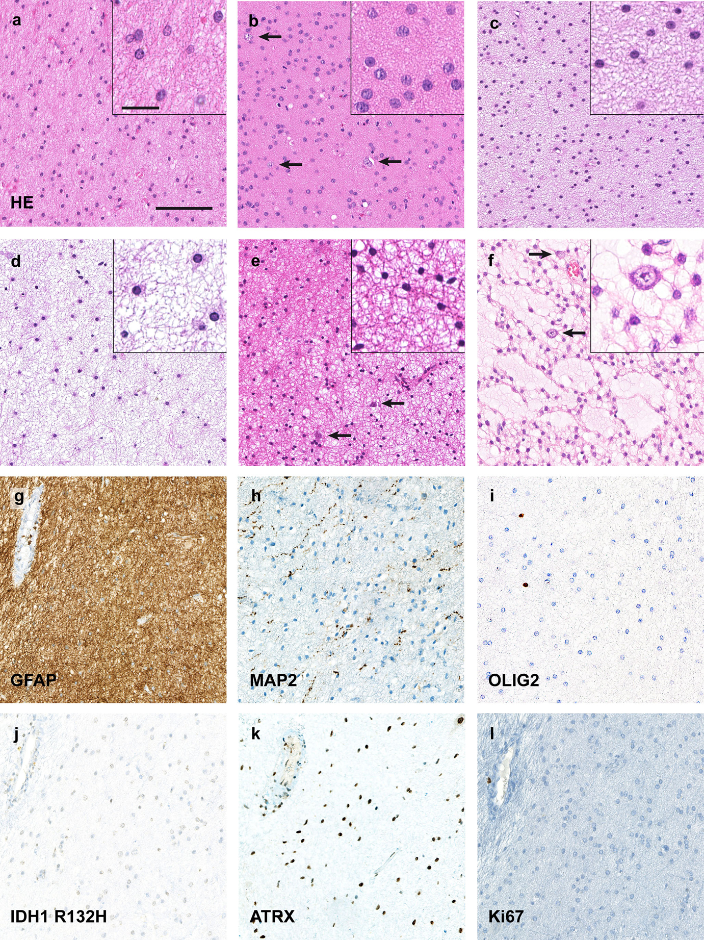
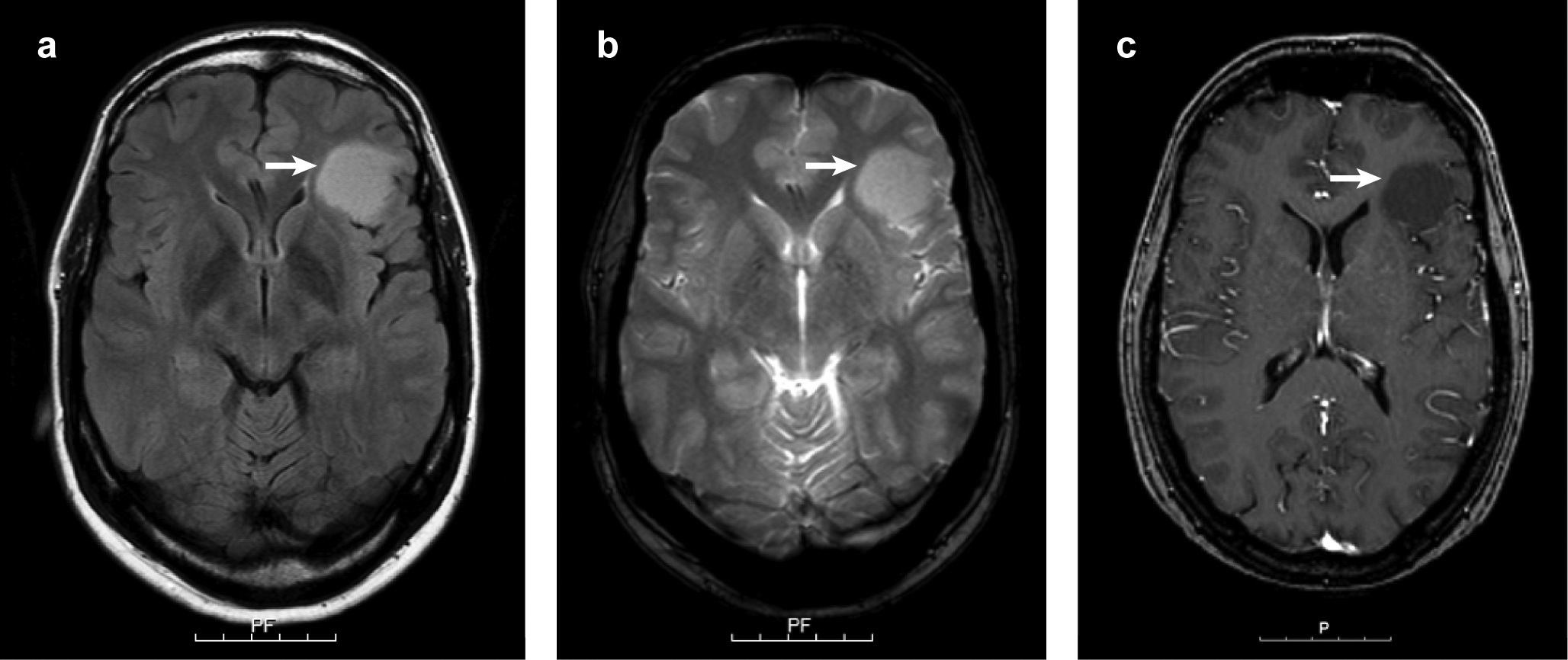
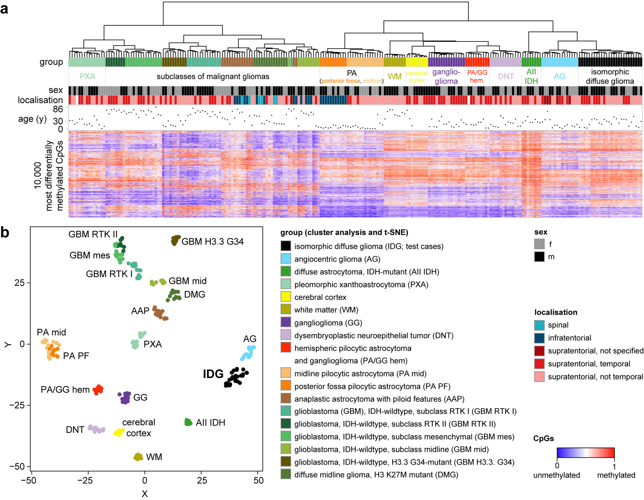
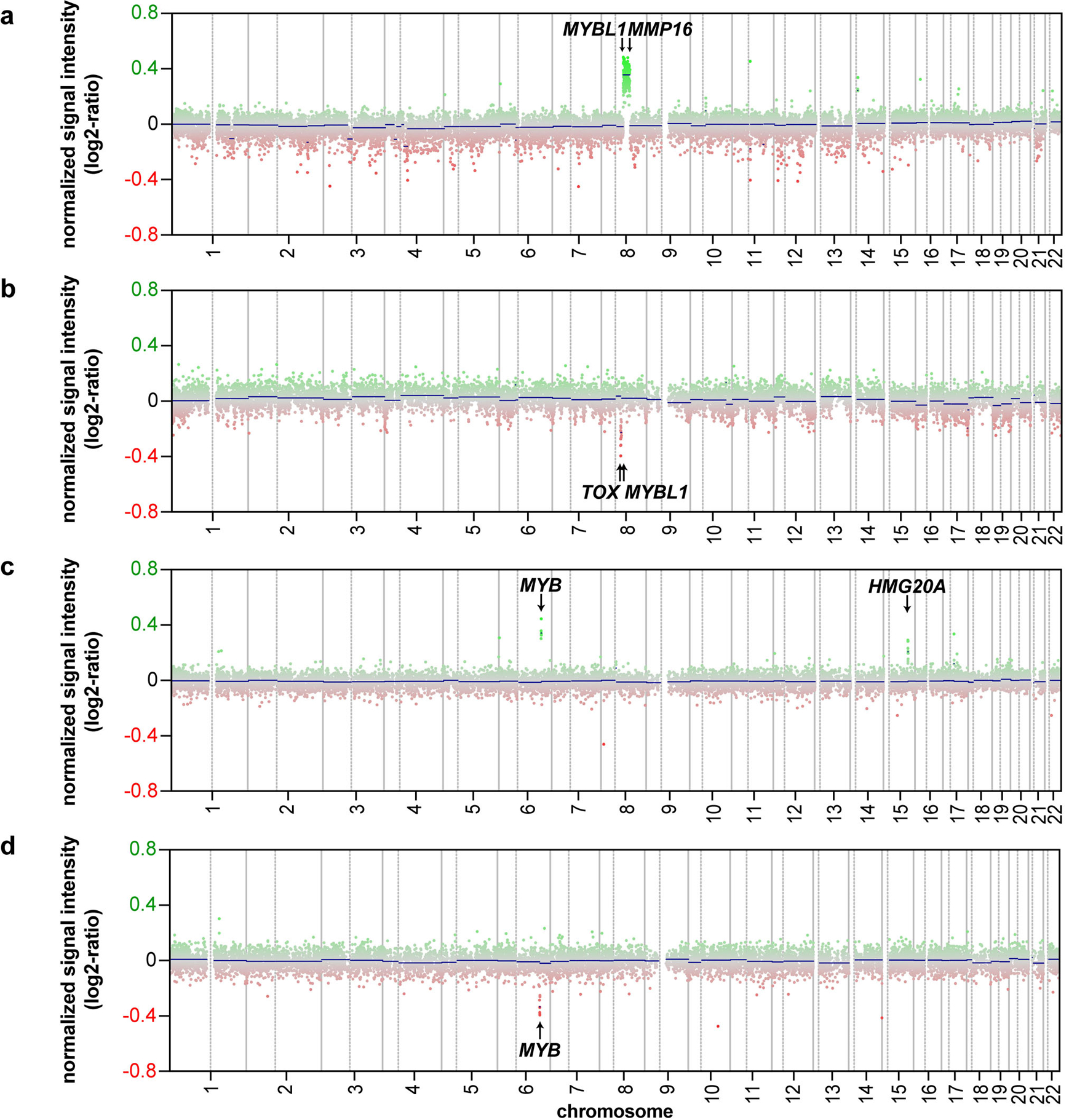
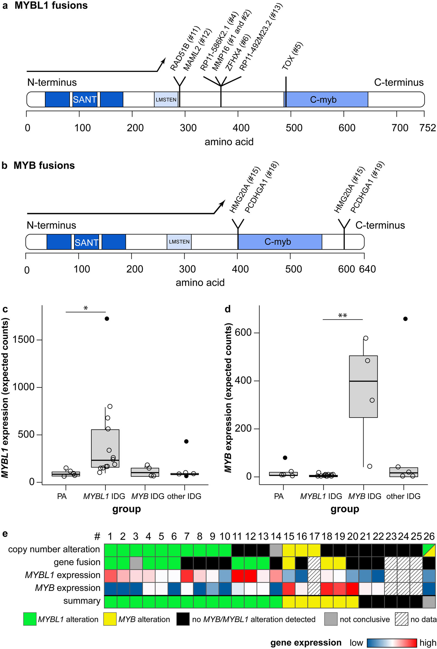
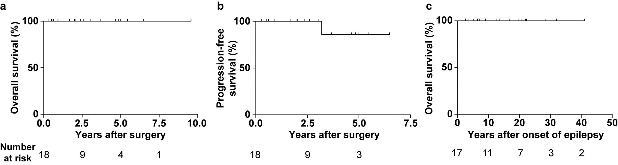
The "isomorphic subtype of diffuse astrocytoma" was identified histologically in 2004 as a supratentorial, highly differentiated glioma with low cellularity, low proliferation and focal diffuse brain infiltration. Patients typically had seizures since childhood and all were operated on as adults. To define the position of these lesions among brain tumours, we histologically, molecularly and clinically analysed 26 histologically prototypical isomorphic diffuse gliomas. Immunohistochemically, they were GFAP-positive, MAP2-, OLIG2- and CD34-negative, nuclear ATRX-expression was retained and proliferation was low. All 24 cases sequenced were IDH-wildtype. In cluster analyses of DNA methylation data, isomorphic diffuse gliomas formed a group clearly distinct from other glial/glio-neuronal brain tumours and normal hemispheric tissue, most closely related to paediatric MYB/MYBL1-altered diffuse astrocytomas and angiocentric gliomas. Half of the isomorphic diffuse gliomas had copy number alterations of MYBL1 or MYB (13/25, 52%). Gene fusions of MYBL1 or MYB with various gene partners were identified in 11/22 (50%) and were associated with an increased RNA-expression of the respective MYB-family gene. Integrating copy number alterations and available RNA sequencing data, 20/26 (77%) of isomorphic diffuse gliomas demonstrated MYBL1 (54%) or MYB (23%) alterations. Clinically, 89% of patients were seizure-free after surgery and all had a good outcome. In summary, we here define a distinct benign tumour class belonging to the family of MYB/MYBL1-altered gliomas. Isomorphic diffuse glioma occurs both in children and adults, has a concise morphology, frequent MYBL1 and MYB alterations and a specific DNA methylation profile. As an exclusively histological diagnosis may be very challenging and as paediatric MYB/MYBL1-altered diffuse astrocytomas may have the same gene fusions, we consider DNA methylation profiling very helpful for their identification.

Keywords: Epilepsy; Gene fusion; Glioma; Isomorphic diffuse glioma; MYB; MYBL1.


Conflict of interest statement

Conflict of interest

D. Capper, D. T. W. Jones, A. von Deimling and S. M. Pfister declare that under the No. EP3067432A1 a patent was applied for a “DNA-methylation based method for classifying tumor species”. D. Capper and A. von Deimling are patent holders of “Methods for the diagnosis and the prognosis of a brain tumor”, the IDH1 R132H-specific antibody used in this manuscript (US 8367347 B2). The patent is under the administrative supervision of the DKFZ technology transfer office.

Figures

Fig. 1
Fig. 1
Isomorphic diffuse gliomas are monomorphic, IDH-wildtype tumours with low proliferation. a–d Histologically typical isomorphic diffuse gliomas showing round, only minimally pleomorphic nuclei, often with speckled chromatin. The cell density is slightly (a, d) to moderately (b, c) increased. The fibrillary matrix between the tumour cells can have slight (c) to extensive (d) microcystic changes. In some tumours scattered pre-existing neurons (arrows in b, e, f) are observed. e Occasionally, isomorphic diffuse gliomas may have smaller, more condensed round nuclei resembling those of oligodendrocytes. f One case focally showed myxoid changes of the matrix with scattered residual neurons, resembling the glio-neuronal element of dysembryoplastic neuroepithelial tumours. In other areas, the tumour also showed a clearly isomorphic growth. g–l Immunohistochemically, isomorphic diffuse gliomas are GFAP-positive (g; same tumour as in a) whereas MAP2 only labels residual neurons and neuronal processes (h). The tumours are OLIG2-negative (i) and IDH1 R132H-negative (j) with a retained nuclear expression of ATRX (k). The Ki67 proliferation index is below 1% (l). Scale bars 200 μm in a for a–l, 25 μm in the inset in a for all insets in a–f
Fig. 2
Fig. 2
Representative MRI images of an isomorphic diffuse glioma. The tumour is hyperintense in FLAIR (arrow in a) and T2-weighted images (arrow in b) while hypointense in the T1-sequence (arrow in c). The tumour did not show contrast enhancement (c). Scale bars 5 cm with 1 cm intervals.
Fig. 3
Fig. 3
Isomorphic diffuse glioma forms a distinct methylation cluster. a Unsupervised hierarchical cluster analysis of 26 isomorphic diffuse gliomas with 246 cases from 17 reference classes. b t-distributed stochastic neighbor embedding (t-SNE) analysis of the same cases. Isomorphic diffuse glioma forms a cluster distinct from other glial/glio-neuronal tumour entities and normal cortex and white matter. The closest relation was found to the cluster of angiocentric glioma.
Fig. 4
Fig. 4
A subset of isomorphic diffuse gliomas has copy number alterations of the MYBL1- and MYB-genes. a–d Exemplary copy number profiles (CNP) of isomorphic diffuse gliomas. Gains are shown in green, losses in red. (a) Gain from MYBL1 to MMP16 (# 2; similar CNP in # 1). (b) Loss from MYBL1 to TOX (# 5). (c) Gain of MYB plus gain of HMG20A on chromosome 15 (# 15). (d) Loss of MYB and the region 3’ of MYB (# 17).
Fig. 5
Fig. 5
Isomorphic diffuse gliomas have fusions of the MYBL1- and MYB-genes and show a corresponding mRNA-overexpression. a, b Canonical MYBL1- (a) and MYB-protein (b) with loci of fusions to different partners. Protein domains in blue: “SANT” DNA-binding domain, “LMSTEN” transactivating domain, “C-myb” C-terminal negative regulatory domain. Fusions result in a deletion of the C-terminal negative regulatory C-myb domain of MYBL1 (a) or MYB (b), or the 3’ miRNA-binding sites of MYB (not included in depiction as no part of the MYB protein). (c, d) Many isomorphic diffuse gliomas show an overexpression of MYBL1 (c) or MYB (d). Compared were pilocytic astrocytomas (“PA”), isomorphic diffuse gliomas with MYBL1-alterations (“MYBL1 IDG”) or MYB-alterations (“MYB IDG”) in the CNP and/or RNA sequencing, and isomorphic diffuse gliomas without evidence of MYBL1/MYB-alterations (“other IDG”). Outliers are depicted with filled circles. p = 0.022 for PA versus MYBL1 IDG in c, p = 0.009 for MYBL1 versus MYB IDG in d (p-values adjusted for multiple comparisons). (e) Summary of alterations of MYBL1 and MYB in the 26 isomorphic diffuse gliomas of this study detected with different methods. For better differentiation, the range of the colour scale for the MYBL1 expression does not account for the outlier. #: case number
Fig. 6
Fig. 6
Patients with isomorphic diffuse gliomas have a good prognosis. (a) Overall survival after surgery of 18 patients with isomorphic diffuse glioma. (b) Progression-free survival after surgery of the same patients. One patient showed a growth of residual tumour and underwent a second surgery three years after the first operation with complete removal of the tumour (recurrence-free since). (c) Overall survival after onset of epilepsy of 17 patients with isomorphic diffuse glioma. Note that no patient with isomorphic diffuse glioma died during follow-up.

References

    1. Bandopadhayay P, Ramkissoon LA, Jain P, Bergthold G, Wala J, Zeid R et al. (2016) MYB-QKI rearrangements in angiocentric glioma drive tumorigenicity through a tripartite mechanism. Nat Genet 48:273–282. doi:10.1038/ng.3500 - DOI - PMC - PubMed
    1. Blumcke I, Aronica E, Urbach H, Alexopoulos A, Gonzalez-Martinez JA (2014) A neuropathology-based approach to epilepsy surgery in brain tumors and proposal for a new terminology use for long-term epilepsy-associated brain tumors. Acta Neuropathol 128:39–54. doi:10.1007/s00401-014-1288-9 - DOI - PMC - PubMed
    1. Blumcke I, Luyken C, Urbach H, Schramm J, Wiestler O (2004) An isomorphic subtype of long-term epilepsy-associated astrocytomas associated with benign prognosis. Acta Neuropathol 107:381–388. doi:10.1007/s00401-004-0833-3 - DOI - PubMed
    1. Blumcke I, Spreafico R, Haaker G, Coras R, Kobow K, Bien CG et al. (2017) Histopathological Findings in Brain Tissue Obtained during Epilepsy Surgery. New Engl J Med 377:1648–1656. doi:10.1056/NEJMoa1703784 - DOI - PubMed
    1. Brat DJ, Verhaak RG, Aldape KD, Yung WK, Salama SR, Cooper LA et al. (2015) Comprehensive, Integrative Genomic Analysis of Diffuse Lower-Grade Gliomas. N Engl J Med 372:2481–2498. doi:10.1056/NEJMoa1402121 - DOI - PMC - PubMed

Publication types