. 2019 Oct 3;105(4):836-843.
doi: 10.1016/j.ajhg.2019.08.008. Epub 2019 Sep 26.

Autosomal-Recessive Mutations in MESD Cause Osteogenesis Imperfecta

Affiliations

Autosomal-Recessive Mutations in MESD Cause Osteogenesis Imperfecta

Shahida Moosa et al. Am J Hum Genet. .

Abstract

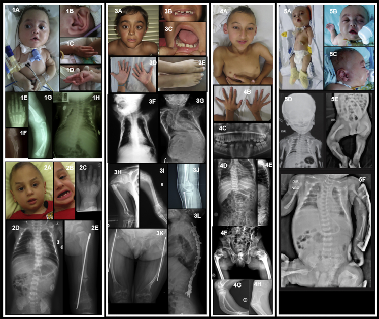
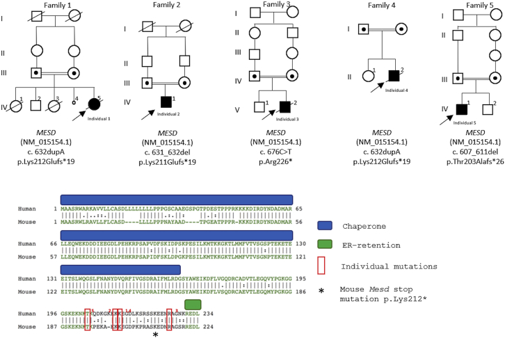
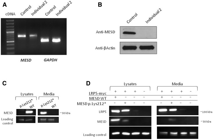
Osteogenesis imperfecta (OI) comprises a genetically heterogeneous group of skeletal fragility diseases. Here, we report on five independent families with a progressively deforming type of OI, in whom we identified four homozygous truncation or frameshift mutations in MESD. Affected individuals had recurrent fractures and at least one had oligodontia. MESD encodes an endoplasmic reticulum (ER) chaperone protein for the canonical Wingless-related integration site (WNT) signaling receptors LRP5 and LRP6. Because complete absence of MESD causes embryonic lethality in mice, we hypothesized that the OI-associated mutations are hypomorphic alleles since these mutations occur downstream of the chaperone activity domain but upstream of ER-retention domain. This would be consistent with the clinical phenotypes of skeletal fragility and oligodontia in persons deficient for LRP5 and LRP6, respectively. When we expressed wild-type (WT) and mutant MESD in HEK293T cells, we detected WT MESD in cell lysate but not in conditioned medium, whereas the converse was true for mutant MESD. We observed that both WT and mutant MESD retained the ability to chaperone LRP5. Thus, OI-associated MESD mutations produce hypomorphic alleles whose failure to remain within the ER significantly reduces but does not completely eliminate LRP5 and LRP6 trafficking. Since these individuals have no eye abnormalities (which occur in individuals completely lacking LRP5) and have neither limb nor brain patterning defects (both of which occur in mice completely lacking LRP6), we infer that bone mass accrual and dental patterning are more sensitive to reduced canonical WNT signaling than are other developmental processes. Biologic agents that can increase LRP5 and LRP6-mediated WNT signaling could benefit individuals with MESD-associated OI.

Keywords: MESD; WNT signaling; osteogenesis imperfecta.


Conflict of interest statement

The authors declare no competing interests.

Figures

Figure 1
Figure 1
Clinical and Radiological Features of Individuals with MESD Mutations (1) Individual 1 from Brazil (Family 1 IV-5) (1A–1B) Facial dysmorphic features include bluish sclerae, mid-face hypoplasia, arched eyebrows, tented upper lip; also note tracheostomy, narrow chest, and rhizomelia; posteriorly rotated ears. (1C–1D) Long fingers with adducted thumbs and contractures of the fifth digits. (1E) Radiograph of left hand showing long, tapering fingers. (1F–1G) Fracture of left ulna and radius at 5 months, healing with callus formation. (1H) Radiograph of chest at 5 months showing bell-shaped, narrow chest with multiple (healing) rib fractures. (2) Individual 2 from Turkey (Family 2 IV-1) (2A–2B) Facial dysmorphic features include tall forehead, arched eyebrows with sparse lateral thirds, micrognathia, posteriorly rotated ears, widely spaced teeth, and oligodontia. (2C) Radiograph of left hand at 8 months showing long, tapering fingers. (2D) Chest radiograph at 5 months showing bell-shaped thorax, thin ribs with healing posterior rib fractures. (2E) Fracture of the left femur at age 4 years, post-surgical rodding. (3) Individual 3 from Portugal (Family 3 V-2) (3A–3C) Facial dysmorphic features include triangular facies, arched eyebrows, left convergent strabismus, midface hypoplasia, thin upper lip, pointed chin, widely spaced teeth, and oligodontia. (3D) Hands showing long fingers with mild contractures. (3E) Feet showing 2-3 partial cutaneous syndactyly with overlapping toes. (3F–3G) Chest radiographs at 6 months and 12 years, respectively, showing multiple rib fractures and progressive scoliosis with vertebral compression fractures. (3H–3I) Upper limb radiographs showing right and left humeral fractures, respectively. (3J) Right knee at age 8 years, showing signs of cyclical pamidronate therapy, rodding of the tibia, and very gracile fibula. (3K) Aged 16 years, thin femurs with bilateral rodding of the femoral shafts post-fracture. (3L) Lateral spinal radiographs at 12 years, showing osteopenia, progressive vertebral compression fractures, and spinal fixation. (4) Individual 4 from Brazil (Family 4 II-2) (4A) He has minimal facial dysmorphic features, slightly upslanting palpebral fissures, and a pointed chin; sclerae are white; skeletal deformity with rhizomelic shortening is noted. (4B) Impression of long fingers with mild interphalangeal contractures. (4C) Panorex radiograph showing disorganized dentition, abnormal premolars, and missing lower jaw teeth (numbers 31, 32, and 41). (4D–4E) Radiographs showing osteopenia, narrow chest, thin ribs with fractures, scoliosis, and vertebral compression fractures. (4F–4H) Radiographs showing skeletal deformity and evidence of fractures of the femoral necks and tibiae. (5) Individual 5 from Brazil (Family 5 IV-1) (5A–5C) Facial dysmorphic features include round face, prominent eyes with long lashes and epicanthic folds, small nose with bulbous tip, tented upper lip, high and narrow palate, retrognathia, and posteriorly rotated ears; also note narrow thorax with inverted nipples, rhizomelic shortening of limbs, and long fingers with contractures. (5D–5F) Radiographs showing generalized osteopenia, multiple fractures (clavicles, ribs, left humerus, femur), narrow thorax, and bowing of femurs bilaterally.
Figure 2
Figure 2
Pedigrees of the Five MESD-Associated OI Individuals, Their MESD Mutations, and the Location of these Mutations in the MESD Protein Upper panel: Family pedigrees and mutations in MESD for each affected individual. Lower panel: Alignment of human MESD with mouse MESD, showing important chaperone activity and ER-retention domains; the first amino acid residues affected by the four different affected individual mutations are noted with red rectangles and the amino acid residue converted to a termination codon in the mouse Mesd expression construct is asterisked.
Figure 3
Figure 3
Individual 2-Derived and Transiently Transfected Cell Cultures Showing That OI-Associated MESD Protein Is Expressed and Capable of Serving as a Chaperone for LRP5, but Unable to Be Retained within Endoplasmic Reticulum of the Cell (A) Agarose gel electrophoretic separation and fluorescence staining of RT-PCR amplimers spanning from exon 1 to exon 3 obtained from wild type (WT, control) and MESD-associated OI (Individual 2) cultured fibroblasts. Spliced MESD amplimers are detected in both samples; spliced GAPDH amplimers serve as a positive control. (B) Immunodetection of MESD following SDS-PAGE in cell lysates from control and Individual 2 fibroblasts. A polyclonal antibody that detects an epitope near the amino-terminal domain of MESD should recognize both WT and mutant MESD. However, only WT MESD is present in the cell lysate. (C) Representative immuno-blot following SDS-PAGE of FLAG epitope-tagged WT or mutant (p.Lys212) MESD in cell lysate and conditioned medium from transiently transfected HEK293T cells. WT MESD is only detected in the cell lysate, whereas mutant MESD is only detected in the conditioned medium. (D) Representative immuno-blots of the same membrane following SDS-PAGE using anti-myc antibody (for LRP5), anti-FLAG antibody (for MESD), anti-beta actin antibody (as a loading control for the cell lysate), and a non-specific cross-reacting band (as a loading control for the conditioned medium) of cell lysate and conditioned medium from HEK293T cells that were transiently transfected with expression vectors for myc-epitope tagged LRP5 (LRP5-myc), FLAG-epitope tagged WT MESD (MESD WT), or FLAG-epitope tagged mutant MESD (MESD p.Lys212). Note co-expression of LRP5-myc with either WT or mutant MESD increases the amount of immuno-detectable LRP5 in cell lysate and in conditioned medium, compared to when LRP5-myc is expressed alone. Also note that mutant MESD becomes detectable at low levels in cell lysate from HEK293T cells when transiently co-expressed with LRP5-myc. Mutant MESD is 13 amino acid residues shorter than WT MESD, which is why it migrates faster during SDS-PAGE.

References

    1. Marini J.C., Forlino A., Bächinger H.P., Bishop N.J., Byers P.H., Paepe A., Fassier F., Fratzl-Zelman N., Kozloff K.M., Krakow D. Osteogenesis imperfecta. Nat. Rev. Dis. Primers. 2017;3:17052. - PubMed
    1. Keupp K., Beleggia F., Kayserili H., Barnes A.M., Steiner M., Semler O., Fischer B., Yigit G., Janda C.Y., Becker J. Mutations in WNT1 cause different forms of bone fragility. Am. J. Hum. Genet. 2013;92:565–574. - PMC - PubMed
    1. Holdener B.C., Faust C., Rosenthal N.S., Magnuson T. msd is required for mesoderm induction in mice. Development. 1994;120:1335–1346. - PubMed
    1. Wines M.E., Shi Y., Lindor M., Holdener B.C. Physical localization of the mesoderm development (mesd) functional region. Genomics. 2000;68:322–329. - PubMed
    1. Hsieh J.C., Lee L., Zhang L., Wefer S., Brown K., DeRossi C., Wines M.E., Rosenquist T., Holdener B.C. Mesd encodes an LRP5/6 chaperone essential for specification of mouse embryonic polarity. Cell. 2003;112:355–367. - PubMed

Publication types

Substances