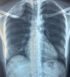
[Benign primitive schwannoma of the pleura]
- PMID: 31565126
- PMCID: PMC6756821
- DOI: 10.11604/pamj.2019.33.164.17625
[Benign primitive schwannoma of the pleura]
Abstract
Schwannoma is a neurogenic tumor originating from Schwann cells. When considering the thoracic region, it is most commonly found in the mediastinum. It commonly appears as a solitary lesion and pleural involvement is extremely rare. We here report the case of a 44-year old woman with benign primitive schwannoma of the pleura whose lesion was detected after radiological assessment for chest pain and dyspnea. The patient underwent complete surgical resection using video-assisted thoracoscopic surgery (VATS) technique. The anatomopathological study showed benign primitive schwannoma of the pleura.
Le schwannome est une tumeur neurogène développée à partir des cellules de Schwann. Dans la région thoracique, le médiastin est le principal site d'apparition du schwannome. Le plus souvent, il s'agit d'une lésion solitaire et la localisation pleurale est extrêmement rare. Nous rapportons un cas de schwannome pleural bénin primitif chez une femme âgée de 44 ans chez qui la lésion a été découverte suite à la réalisation d'un bilan radiologique pour une douleur thoracique et une dyspnée. Le patient a eu résection chirurgicale complète de cette tumeur sous vidéo thoracoscopie. L'étude anatomopathologique a conclu à un schwannome bénin primitif de la plèvre.
Keywords: CT scan; Schwannoma; pleura; video-assisted thoracoscopic surgery (VATS).
© Rachid Marouf et al.
Conflict of interest statement
Les auteurs ne déclarent aucun conflit d'intérêts.
Figures


References
-
- Izzillo R, Lopez I, Perret C, Badaro D, Busy F. Volumineux schwannome bénin pleural. J Radiol. 1999;80(8):866–8. - PubMed
-
- Gabriel R, Rao A. Digital neurilemmoma: a case report. Bahrain Med Bull. 2002;24(2):76–7.
-
- Mc Clenathan JH, Bloom RJ. Peripheral tumors of the intercostal nerves. Ann Thorac Surg. 2004;78(2):713–4. - PubMed
-
- Athanassiadi K, Kalavrouziotis G, Rondogianni D, Loutsidis A, Hatzimichalis A, Bellenis I. Primary chest wall tumors: early and long-term results of surgical treatment. Eur J Cardiothorac Surg. 2001 May;19(5):589–93. - PubMed
Publication types
MeSH terms
LinkOut - more resources
Full Text Sources
