. 2018 Dec 3;3(3):1800058.
doi: 10.1002/gch2.201800058. eCollection 2019 Mar.

Vanadium Dioxide Nanocoating Induces Tumor Cell Death through Mitochondrial Electron Transport Chain Interruption

Affiliations

Vanadium Dioxide Nanocoating Induces Tumor Cell Death through Mitochondrial Electron Transport Chain Interruption

Jinhua Li et al. Glob Chall. .

Abstract

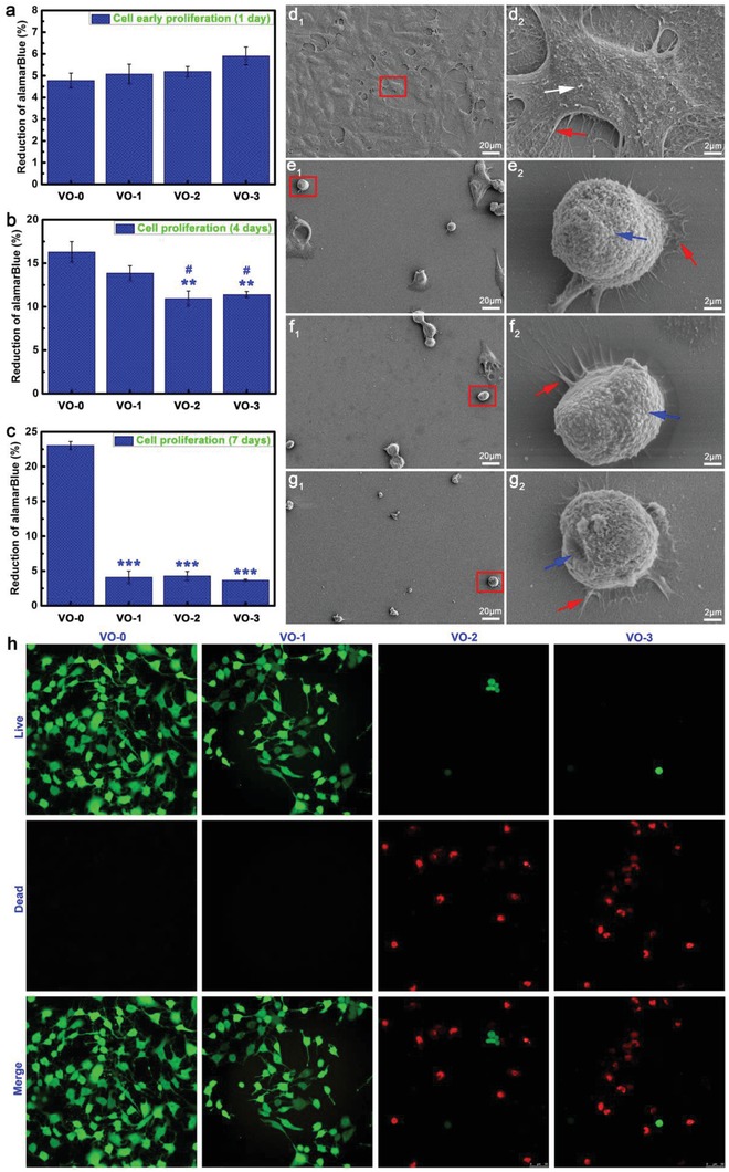
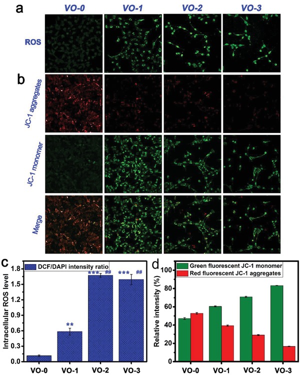
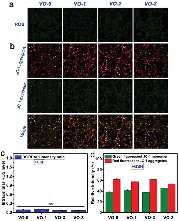
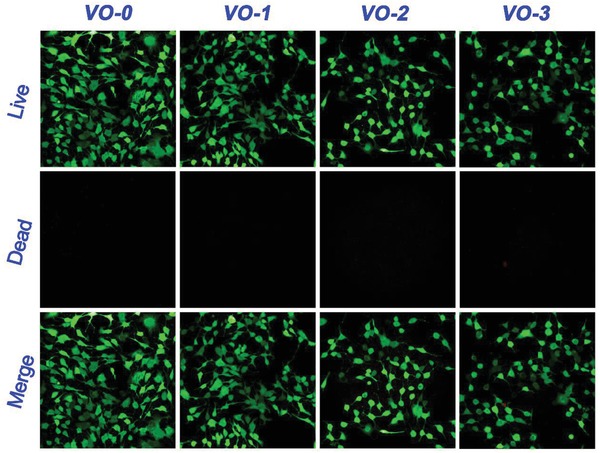
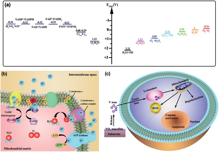
A biomaterials surface enabling the induction of tumor cell death is particularly desirable for implantable biomedical devices that directly contact tumor tissues. However, this specific antitumor feature is rarely found. Consequently, an antitumor-cell nanocoating comprised of vanadium dioxide (VO2) prepared by customized reactive magnetron sputtering has been proposed, and its antitumor-growth capability has been demonstrated using human cholangiocarcinoma cells. The results reveal that the VO2 nanocoating is able to interrupt the mitochondrial electron transport chain and then elevate the intracellular reactive oxygen species levels, leading to the collapse of the mitochondrial membrane potential and the destruction of cell redox homeostasis. Indeed, this chain reaction can effectively trigger oxidative damage in the cholangiocarcinoma cells. Additionally, this study has provided new insights into designing a tumor-cell-inhibited biomaterial surface, which is modulated by the mechanism of mitochondria-targeting tumor cell death.

Keywords: anticancer; charge transfer; functional coatings and films; mitochondria; vanadium dioxide.


Conflict of interest statement

The authors declare no conflict of interest.

Figures

Figure 1
Figure 1
a) Schematic illustration for the deposition of VO2 nanofilm on substrate synthesized by reactive sputtering of metallic V target with O2 gas introduction under a heating condition. AFM images of the surfaces of b) VO‐1, c) VO‐2, and d) VO‐3 samples. e) XRD patterns of the samples VO‐0, VO‐1, VO‐2, and VO‐3.
Figure 2
Figure 2
a) Measurement results of surface zeta potentials of samples VO‐0, VO‐1, VO‐2, and VO‐3. b) Chemical valence states of V element before and after soaking in PBS for 1 and 14 days. c) Release profiles of vanadium ions from samples VO‐0, VO‐1, VO‐2, and VO‐3 within 14 days.
Figure 3
Figure 3
Tumor cell viability results after a) 1 day, b) 4 days, and c) 7 days of culture on the samples VO‐0, VO‐1, VO‐2, and VO‐3 (Note: **P < 0.01, ***P < 0.001 versus VO‐0; # P < 0.05 versus VO‐1). Tumor cell morphology and cell–material interactions after 4 days of culture on the samples d1,d2) VO‐0, e1,e2) VO‐1, f1,f2) VO‐2, and g1,g2) VO‐3 (Note: white arrow, indicating the exuberant extracellular matrix mineralization; red arrow, indicating the abundant lamellipodia and filopodia extensions; blue arrow, indicating the deteriorating cell membrane). h) Live/dead fluorescence staining results of tumor cells after 4 days of culture on the samples VO‐0, VO‐1, VO‐2, and VO‐3.
Figure 4
Figure 4
Fluorescence staining images of a) intracellular ROS for tumor cells and b) mitochondria membrane potentials after 4 days of culture on the samples VO‐0, VO‐1, VO‐2, and VO‐3, accompanied by the corresponding quantitative results of c) ROS levels and d) membrane potentials. Note: **P < 0.01, ***P < 0.001 versus VO‐0; ## P < 0.01 versus VO‐1.
Figure 5
Figure 5
Fluorescence staining images of a) intracellular ROS for tumor cells and b) mitochondria membrane potentials after 4 days of culture on the samples VO‐0, VO‐1, VO‐2, and VO‐3 with adding GSH, accompanied by the corresponding quantitative results of c) ROS levels and d) membrane potentials. Note: ns, not significant.
Figure 6
Figure 6
Live/dead fluorescence staining results of tumor cells after 4 days of culture on the samples VO‐0, VO‐1, VO‐2, and VO‐3 with adding GSH.
Scheme 1
Scheme 1
Schematic illustration of anticancer mechanism of VO2 nanocoating against tumor cells: a) Redox potentials for the redox couples in mitochondrial electron transfer chain and ROS‐associated redox couples, indicating the feasibility of mitochondrial electron transport chain interruption and the availability of intracellular ROS production. b) Diagram of the mitochondrial electron transport chain and associated interactions, in which electron leakage is able to elevate intracellular ROS production, thereby leading to mitochondria dysfunction. c) Mitochondria‐targeting anticancer process of the VO2 nanocoating platform by releasing vanadate ions to cause mitochondria depolarization and oxidative damage, thereby resulting in tumor cell apoptosis.

Similar articles

Cited by

References

    1. a) Hanker J., Giammara B., Science 1988, 242, 885; - PubMed
    2. b) Tang R., Moyano D. F., Subramani C., Yan B., Jeoung E., Tonga G. Y., Duncan B., Yeh Y.‐C., Jiang Z., Kim C., Rotello V. M., Adv. Mater. 2014, 26, 3310; - PMC - PubMed
    3. c) Bowen P. K., Drelich J., Goldman J., Adv. Mater. 2013, 25, 2577; - PubMed
    4. d) Wang H., Kwok D. T. K., Xu M., Shi H., Wu Z., Zhang W., Chu P. K., Adv. Mater. 2012, 24, 3315; - PubMed
    5. e) Banerjee I., Pangule R. C., Kane R. S., Adv. Mater. 2011, 23, 690; - PubMed
    6. f) Hu Y., Cai K. Y., Luo Z., Jandt K. D., Adv. Mater. 2010, 22, 4146. - PubMed
    1. a) Weilbaecher K. N., Guise T. A., McCauley L. K., Nat. Rev. Cancer 2011, 11, 411; - PMC - PubMed
    2. b) Luetke A., Meyers P. A., Lewis I., Juergens H., Cancer Treat. Rev. 2014, 40, 523. - PubMed
    1. a) Ma H., Luo J., Sun Z., Xia L., Shi M., Liu M., Chang J., Wu C., Biomaterials 2016, 111, 138; - PubMed
    2. b) Zhang M., Cheng H., Gong Z., Zhang J., Liu X., Wang B., Ban L., Zeng Y., Zhu Z., Adv. Funct. Mater. 2017, 27, 1703932.
    1. a) Ma H., Jiang C., Zhai D., Luo Y., Chen Y., Lv F., Yi Z., Deng Y., Wang J., Chang J., Wu C., Adv. Funct. Mater. 2016, 26, 1197;
    2. b) Wang X., Li T., Ma H., Zhai D., Jiang C., Chang J., Wang J., Wu C., NPG Asia Mater. 2017, 9, e376.
    1. a) Khan S. A., Thomas H. C., Davidson B. R., Taylor‐Robinson S. D., Lancet 2005, 366, 1303; - PubMed
    2. b) Wang A., Wu L., Lin J., Han L., Bian J., Wu Y., Robson S. C., Xue L., Ge Y., Sang X., Wang W., Zhao H., Nat. Commun. 2018, 9, 894. - PMC - PubMed

LinkOut - more resources