

Quantifying skin photodamage with spatial frequency domain imaging: statistical results
- PMID: 31565518
- PMCID: PMC6757479
- DOI: 10.1364/BOE.10.004676
Quantifying skin photodamage with spatial frequency domain imaging: statistical results
Abstract
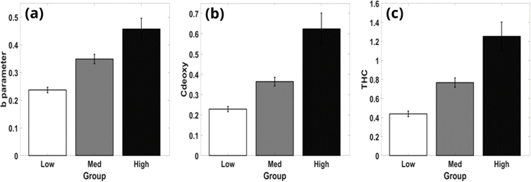
We investigated the change in optical properties and vascular parameters to characterize skin tissue from mild photodamage to actinic keratosis (AK) with comparison to a published photodamage scale. Multi-wavelength spatial frequency domain imaging (SFDI) measurements were performed on the dorsal forearms of 55 adult subjects with various amounts of photodamage. Dermatologists rated the levels of photodamage based upon the photographs in blinded fashion to allow comparison with SFDI data. For characterization of statistical data, we used artificial neural networks. Our results indicate that optical and vascular parameters can be used to quantify photodamage and can discriminate between the stages as low, medium, and high grades, with the best performance of ∼70%, ∼76% and 80% for characterization of low- medium- and high-grade lesions, respectively. Ultimately, clinicians can use this noninvasive approach for risk assessment and frequent monitoring of high-risk populations.
© 2019 Optical Society of America under the terms of the OSA Open Access Publishing Agreement.
Conflict of interest statement
The authors declare that there are no conflicts of interest related to this article.
Figures
Similar articles
-
Inter- and Intra-physician variation in quantifying actinic keratosis skin photodamage.J Clin Investig Dermatol. 2020 Sep;8(2):4. Epub 2020 Sep 6. J Clin Investig Dermatol. 2020. PMID: 33088904 Free PMC article.
-
Noninvasive mesoscopic imaging of actinic skin damage using spatial frequency domain imaging.Biomed Opt Express. 2017 May 23;8(6):3045-3052. doi: 10.1364/BOE.8.003045. eCollection 2017 Jun 1. Biomed Opt Express. 2017. PMID: 28663925 Free PMC article.
-
Expert Consensus on Cosmetic Outcomes After Treatment of Actinic Keratosis.J Drugs Dermatol. 2017 Mar 1;16(3):260-264. J Drugs Dermatol. 2017. PMID: 28301622
-
Early assessment of burn severity in human tissue ex vivo with multi-wavelength spatial frequency domain imaging.Toxicol In Vitro. 2018 Oct;52:251-254. doi: 10.1016/j.tiv.2018.05.015. Epub 2018 Jun 1. Toxicol In Vitro. 2018. PMID: 29859991 Free PMC article.
-
Management of actinic keratosis at specific body sites in patients at high risk of carcinoma lesions: expert consensus from the AKTeam™ of expert clinicians.J Eur Acad Dermatol Venereol. 2018 Mar;32(3):339-346. doi: 10.1111/jdv.14753. Epub 2018 Jan 15. J Eur Acad Dermatol Venereol. 2018. PMID: 29235161
Cited by
-
Inter- and Intra-physician variation in quantifying actinic keratosis skin photodamage.J Clin Investig Dermatol. 2020 Sep;8(2):4. Epub 2020 Sep 6. J Clin Investig Dermatol. 2020. PMID: 33088904 Free PMC article.
-
Mapping cutaneous field carcinogenesis of nonmelanoma skin cancer using mesoscopic imaging of pro-inflammation cues.Exp Dermatol. 2024 Apr;33(4):e15076. doi: 10.1111/exd.15076. Exp Dermatol. 2024. PMID: 38610095 Free PMC article. Review.
-
Multimodal hyperspectral fluorescence and spatial frequency domain imaging for tissue health diagnostics of the oral cavity.Biomed Opt Express. 2021 Oct 18;12(11):6954-6968. doi: 10.1364/BOE.439663. eCollection 2021 Nov 1. Biomed Opt Express. 2021. PMID: 34858691 Free PMC article.
-
Dual-DMD hyperspectral spatial frequency domain imaging (SFDI) using dispersed broadband illumination with a demonstration of blood stain spectral monitoring.Biomed Opt Express. 2020 Dec 24;12(1):676-688. doi: 10.1364/BOE.411976. eCollection 2021 Jan 1. Biomed Opt Express. 2020. PMID: 33520393 Free PMC article.
-
Spatial Frequency Domain Imaging System Calibration, Correction and Application for Pear Surface Damage Detection.Foods. 2021 Sep 11;10(9):2151. doi: 10.3390/foods10092151. Foods. 2021. PMID: 34574261 Free PMC article.
References
-
- Konger R. L., Xu Z., Sahu R. P., Rashid B. M., Mehta S. R., Mohamed D. R., DaSilva-Arnold S. C., Bradish J. R., Warren S. J., Kim Y. L., “Spatiotemporal assessments of dermal hyperemia enable accurate prediction of experimental cutaneous carcinogenesis as well as chemopreventive activity,” Cancer Res. 73(1), 150–159 (2013).10.1158/0008-5472.CAN-12-2670 - DOI - PMC - PubMed
LinkOut - more resources
Full Text Sources