

Phage constructs targeting gonadotropin-releasing hormone for fertility control: evaluation in cats
- PMID: 31566070
- PMCID: PMC10814494
- DOI: 10.1177/1098612X19875831
Phage constructs targeting gonadotropin-releasing hormone for fertility control: evaluation in cats
Abstract
Objectives: Phage-gonadotropin-releasing hormone (GnRH) constructs with potential contraceptive properties were generated in our previous study via selection from a phage display library using neutralizing GnRH antibodies as selection targets. In mice, these constructs invoked the production of antibodies against GnRH and suppressed serum testosterone. The goal of this study was to evaluate this vaccine against GnRH for its potential to suppress reproductive characteristics in cats.
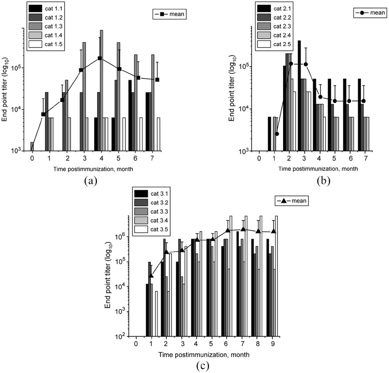
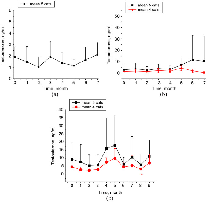
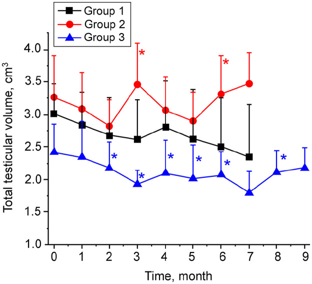
Methods: Sexually mature male cats were injected with a phage-GnRH vaccine using the following treatment groups: (1) single phage-GnRH vaccine with adjuvant; (2) phage-GnRH vaccine without adjuvant and half-dose booster 1 month later; or (3) phage-GnRH vaccine with adjuvant and two half-dose boosters with adjuvant 3 and 6 months later. Anti-GnRH antibodies and serum testosterone, testicular volume and sperm characteristics were evaluated monthly for 7-9 months.
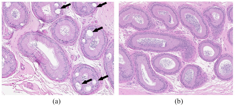
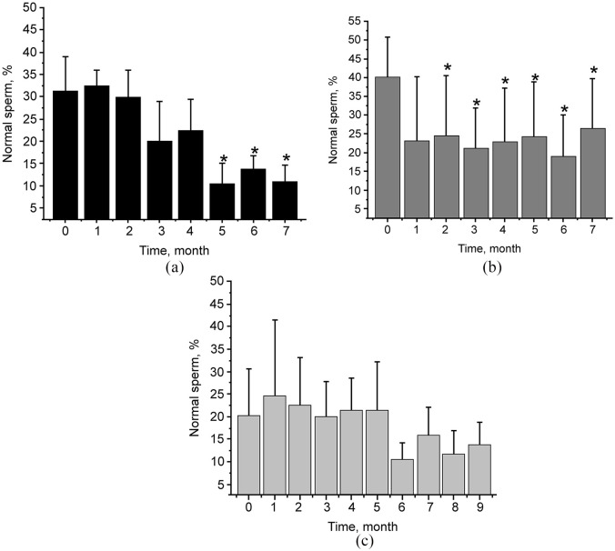
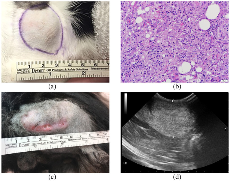
Results: All cats developed anti-GnRH antibodies following immunization. Serum antibody titers increased significantly after booster immunizations. In group 3, serum testosterone was suppressed 8 months after primary immunization. Total testicular volume decreased in group 1 by 24-42% and in group 3 by 15-36% at 7 months after immunization, indicating potential gonadal atrophy. Vacuolation of epididymides was observed histologically. Although all cats produced sperm at the conclusion of the study, normal morphology was decreased as much as 38%. Phage alone produced no local or systemic reactions. Immunization of phage with AdjuVac produced unacceptable injection site reactions.
Conclusions and relevance: Our phage-based vaccine against GnRH demonstrated a potential for fertility impairment in cats. Future research is required to optimize vaccine regimens and identify animal age groups most responsive to the vaccine. If permanent contraception (highly desirable in feral and shelter cats) cannot be achieved, the vaccine has a potential use in zoo animals or pets where multiple administrations are more practical and/or reversible infertility is desirable.
Keywords: Fertility control; GnRH; filamentous phage; gonadotropin-releasing hormone.
Conflict of interest statement
The authors declared no potential conflicts of interest with respect to the research, authorship, and/or publication of this article.
Figures

Similar articles
-
Humoral immune responses against gonadotropin releasing hormone elicited by immunization with phage-peptide constructs obtained via phage display.J Biotechnol. 2015 Dec 20;216:20-8. doi: 10.1016/j.jbiotec.2015.10.001. Epub 2015 Oct 9. J Biotechnol. 2015. PMID: 26456116
-
Generation and characterization of phage-GnRH chemical conjugates for potential use in cat and dog immunocontraception.Reprod Domest Anim. 2012 Dec;47 Suppl 6:406-11. doi: 10.1111/rda.12061. Reprod Domest Anim. 2012. PMID: 23279551
-
Long-term fertility control in female cats with GonaCon™, a GnRH immunocontraceptive.Theriogenology. 2011 Nov;76(8):1517-25. doi: 10.1016/j.theriogenology.2011.06.022. Epub 2011 Aug 10. Theriogenology. 2011. PMID: 21835455 Clinical Trial.
-
Vaccines for control of fertility and hormone dependent cancers.Int J Immunopharmacol. 1992 Apr;14(3):511-4. doi: 10.1016/0192-0561(92)90183-l. Int J Immunopharmacol. 1992. PMID: 1618603 Review.
-
Vaccines for feline contraception: GonaCon GnRH-hemocyanin conjugate immunocontraceptive.J Feline Med Surg. 2015 Sep;17(9):758-65. doi: 10.1177/1098612X15594989. J Feline Med Surg. 2015. PMID: 26323799 Free PMC article. Review.
Cited by
-
Aspects of Phage-Based Vaccines for Protein and Epitope Immunization.Vaccines (Basel). 2023 Feb 14;11(2):436. doi: 10.3390/vaccines11020436. Vaccines (Basel). 2023. PMID: 36851313 Free PMC article. Review.
-
Durable contraception in the female domestic cat using viral-vectored delivery of a feline anti-Müllerian hormone transgene.Nat Commun. 2023 Jun 6;14(1):3140. doi: 10.1038/s41467-023-38721-0. Nat Commun. 2023. PMID: 37280258 Free PMC article.
-
Bacteriophages as Potential Tools for Use in Antimicrobial Therapy and Vaccine Development.Pharmaceuticals (Basel). 2021 Apr 5;14(4):331. doi: 10.3390/ph14040331. Pharmaceuticals (Basel). 2021. PMID: 33916345 Free PMC article. Review.
-
Comprehensive Evaluation and Future Perspectives of Non-Surgical Contraceptive Methods in Female Cats and Dogs.Animals (Basel). 2025 May 21;15(10):1501. doi: 10.3390/ani15101501. Animals (Basel). 2025. PMID: 40427377 Free PMC article. Review.
-
Epigenetic repression of gonadotropin gene expression via a GnRH-mediated DNA delivery system.Gene Ther. 2022 May;29(5):294-303. doi: 10.1038/s41434-022-00325-6. Epub 2022 Mar 17. Gene Ther. 2022. PMID: 35301447
References
-
- Goericke-Pesch S, Wehrend A, Georgiev P. Suppression of fertility in adult cats. Reprod Domest Anim 2014; 49 Suppl 2: 33–40. - PubMed
-
- Vansandt LM, Kutzler MA, Fischer AE, et al.. Safety and effectiveness of a single and repeat intramuscular injection of a GnRH vaccine (GonaCon) in adult female domestic cats. Reprod Domest Anim 2017; 52 Suppl 2: 348–353. - PubMed
-
- Gupta SK, Minhas V. Wildlife population management: are contraceptive vaccines a feasible proposition? Front Biosci 2017; 9: 357–374. - PubMed
Publication types
MeSH terms
Substances
LinkOut - more resources
Full Text Sources
Medical
Miscellaneous
