. 2020 May 1;116(6):1161-1174.
doi: 10.1093/cvr/cvz240.

Impaired NF-κB signalling underlies cyclophilin D-mediated mitochondrial permeability transition pore opening in doxorubicin cardiomyopathy

Affiliations

Impaired NF-κB signalling underlies cyclophilin D-mediated mitochondrial permeability transition pore opening in doxorubicin cardiomyopathy

Rimpy Dhingra et al. Cardiovasc Res. .

Abstract

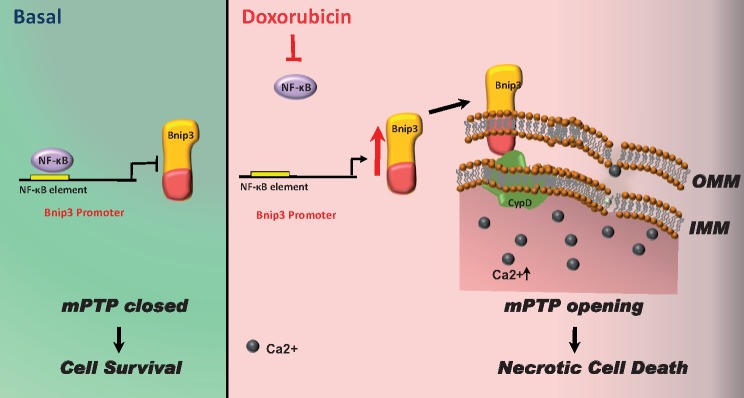
Aims: The chemotherapy drug doxorubicin (Dox) is commonly used for treating a variety of human cancers; however, it is highly cardiotoxic and induces heart failure. We previously reported that the Bcl-2 mitochondrial death protein Bcl-2/19kDa interaction protein 3 (Bnip3), is critical for provoking mitochondrial perturbations and necrotic cell death in response to Dox; however, the underlying mechanisms had not been elucidated. Herein, we investigated mechanism that drives Bnip3 gene activation and downstream effectors of Bnip3-mediated mitochondrial perturbations and cell death in cardiac myocytes treated with Dox.

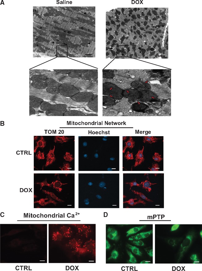
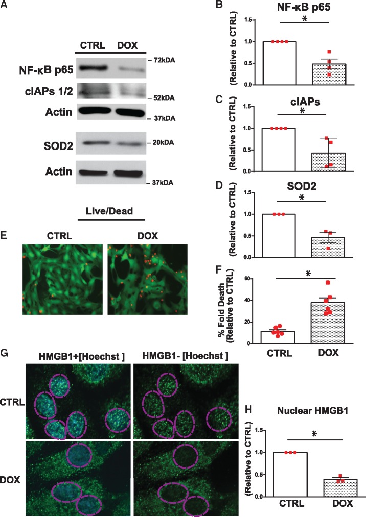
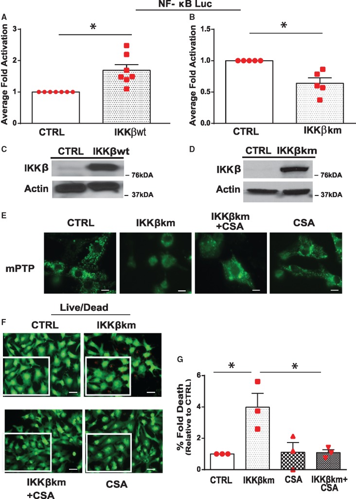
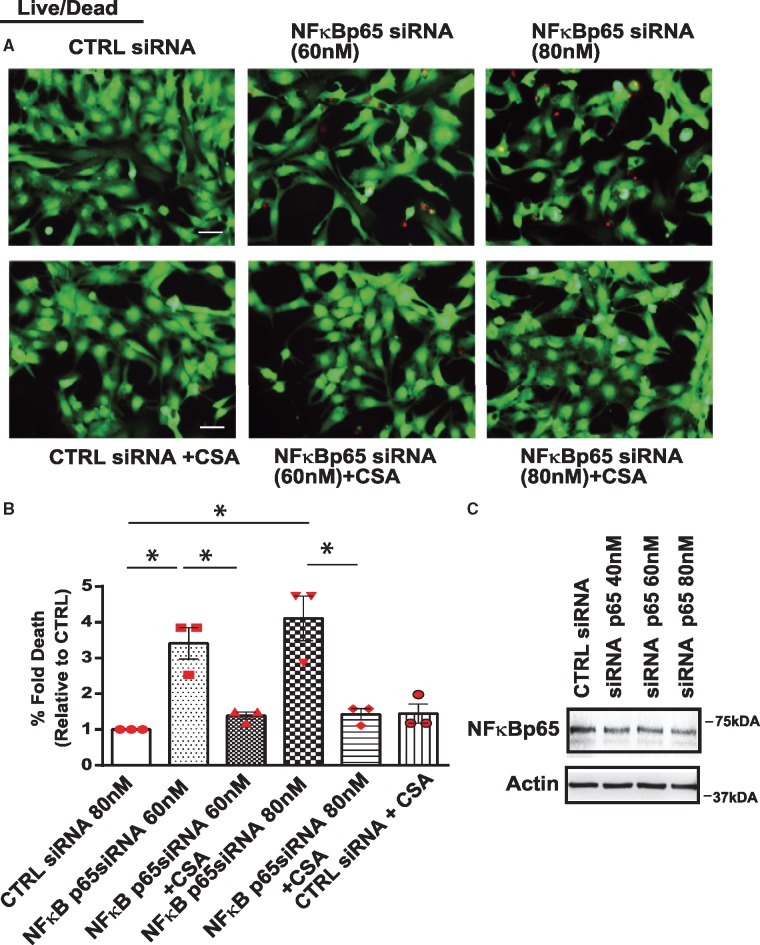
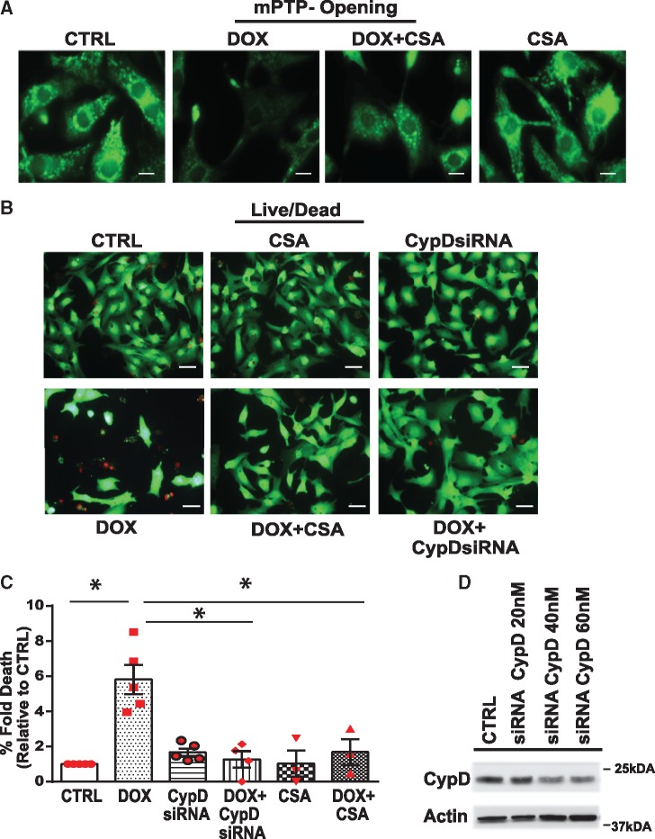
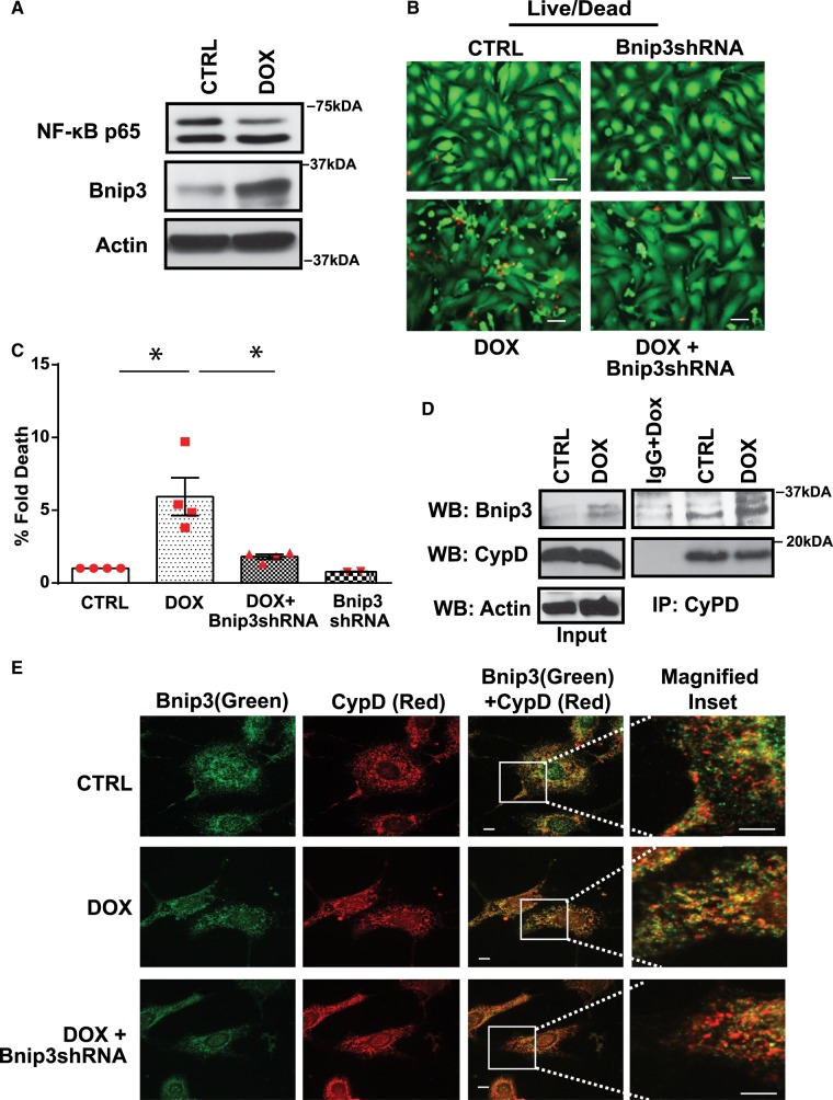
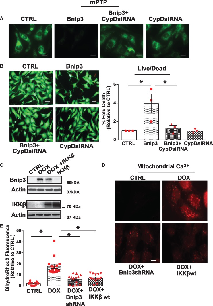
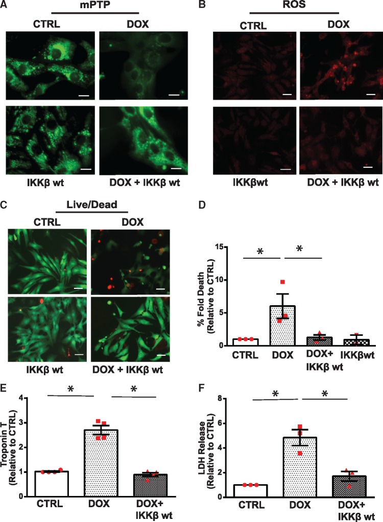
Methods and results: Nuclear factor-κB (NF-κB) signalling, which transcriptionally silences Bnip3 activation under basal states in cardiac myocytes was dramatically reduced following Dox treatment. This was accompanied by Bnip3 gene activation, mitochondrial injury including calcium influx, permeability transition pore (mPTP) opening, loss of nuclear high mobility group protein 1, reactive oxygen species production, and cell death. Interestingly, impaired NF-κB signalling in cells treated with Dox was accompanied by protein complexes between Bnip3 and cyclophilin D (CypD). Notably, Bnip3-mediated mPTP opening was suppressed by inhibition of CypD-demonstrating that CypD functionally operates downstream of Bnip3. Moreover, restoring IKKβ-NF-κB activity in cardiac myocytes treated with Dox suppressed Bnip3 expression, mitochondrial perturbations, and necrotic cell death.

Conclusions: The findings of the present study reveal a novel signalling pathway that functionally couples NF-κB and Dox cardiomyopathy to a mechanism that is mutually dependent upon and obligatorily linked to the transcriptional control of Bnip3. Our findings further demonstrate that mitochondrial injury and necrotic cell death induced by Bnip3 is contingent upon CypD. Hence, maintaining NF-κB signalling may prove beneficial in reducing mitochondrial dysfunction and heart failure in cancer patients undergoing Dox chemotherapy.

Keywords: Bnip3; Cardiac myocytes; Cell death; Doxorubicin; Mitochondria; NF-κB.


Figures

Graphical Abstract
Graphical Abstract
Figure 1
Figure 1
Dox provokes morphological and functional defects to mitochondria. Mitochondrial morphology in mice treated with saline (CTRL) or Dox. (A) Representative electron micrograph (5800× magnification) of heart muscle derived from saline (0.9%) and Dox (20 mg/kg) treated mice, with magnified insets below, scale bar 2 µm. (Left panel) Saline-treated mice exhibit normal mitochondrial morphology. (Right panel) Dox-treated mice displaying abnormal mitochondria with disrupted cristae and electron dense granules consistent with calcium deposits (red arrows). (B) Mitochondrial morphology assessed by epifluorescence microscopy using mitochondrial outer membrane protein Tom 20 (Red) and nuclear DNA (blue, Hoechst 33258), scale bar 10 µm. (C) Representative images of mitochondrial calcium uptake by Rhod-2AM (red fluorescence, see Section 2 for details), scale bar 10 µm. (D) Mitochondrial permeability transition pore (mPTP) opening assessed by using Calcein-AM in the presence of cobalt chloride, loss of green fluorescence indicates mPTP opening, scale bar 10 µm.
Figure 2
Figure 2
Dox impairs NF-κB signalling and promotes necrotic cell death. NF-κB signalling and cell viability was determined in saline (CTRL) and Dox-treated cardiomyocytes. (A) Representative WBs of cell lysate derived from CTRL and Dox-treated cardiomyocytes. The filter was probed with antibodies directed against p65 subunit of NF-κB and its transcriptional targets, cIAPS1/2, and SOD2. Actin served as a protein loading control. (B–D) Quantitative analysis of proteins normalized to actin. Statistical significance determined using Student’s t-test for NF-κB, Dox vs. CTRL *P = 0.004; cIAPs, Dox vs. CTRL *P = 0.015 and SOD2, Dox vs. CTRL *P = 0.012. (E) Representative images of CTRL and Dox-treated cells assessed for cell viability with Calcein-AM and ethidium homodimer for detection of live (green) and dead (red) cells, respectively, scale bar 40µm. (F) Histogram depicts quantitative data for E. Data are expressed as mean ± SEM derived from at least n = 6 independent cardiomyocyte isolations. Unpaired two-tailed Student’s t-test was used to compare mean differences between groups. Statistically significant difference of Dox condition from control, *P = 0.002. (G) High mobility group box 1 immunostaining (HMGB1, green fluorescence) as an index of necrosis. Nuclear DNA stained with Hoechst 33528 (blue fluorescence). Dotted circle demarks nucleus, scale bar 10 µm. (H) Quantitative analysis of nuclear HMGB1 shown in G. Data are expressed as mean ± SEM derived from n = 3 independent cardiomyocyte isolations. Unpaired two tailed Student’s t-test was used to compare mean differences between groups. Statistically significant difference of Dox vs. CTRL *P = 0.0001.
Figure 3
Figure 3
Inhibition of cyclophilin D suppresses cell death of cardiac myocytes expressing IKKβK-M. (A, B) NF-κB gene transcription was assessed by luciferase promoter reporter assay in 293 cells in the presence of IKKβwt (A) or kinase defective form of IKKβ (IKKβK-M) in B. Data were normalized to CTRL and presented as mean ± SEM derived from at least n = 3 experiments. Unpaired two-tailed Student’s t-test was used to compare mean differences between groups. Statistical significance between CTRL and IKKβwt, *P = 0.0026; between CTRL and IKKβK-M, *P = 0.003. (C, D) Expression of IKKβwt and IKKβK-M. (E–G) mPTP opening (C, scale bar 10 µm) and cell viability (scale bar 40 µm, D, E) of ventricular myocytes expressing IKKβK-M in the absence and presence of Cyclosporine A (CSA, 2μM, inhibitor of Cyclophilin D). mPTP opening and cell viability were assessed by epifluorescence microscopy (see Section 2 for details). Data are expressed as mean ± SEM (n = 3 experiments). Statistical significance between the groups was analysed by one-way ANOVA and Bonferroni post hoc test; IKKβK-M vs. CTRL,*P = 0.0145; IKKβK-M vs. IKKβK-M + CSA, *P = 0.0170; IKKβK-M vs. CSA, *P = 0.0177.
Figure 4
Figure 4
Inhibition of cyclophilin D suppresses cell death in cardiac myocytes following NF-κB knock-down. Cardiomyocytes expressing scrambled siRNA or siRNA directed against NF-κB p65 (60 nM and 80 nM) assessed for cell viability assay in the absence and presence of CSA. (A) Representative images for cell viability, scale bar 40 µm. (B) Histogram shows quantitative data for A. Data are expressed as mean ± SEM derived from n = 3 independent experiments. Statistical significance between the groups marked in the histogram, analysed by one-way ANOVA and Bonferroni post hoc test, CTRL vs. NF-κB p65si 60 nM *P = 0.0015; CTRL vs. NF-κB p65si 80 nM *P = 0.0002; NF-κB p65si 60 nM vs. NF-κB p65si 60 nM+CSA *P = 0.006; NF-κB p65si 80 nM vs. NF-κB p65si 80 nM+CSA*P = 0.0006. (C) WB analysis of the cell lysate derived from cells expressing siRNA directed against NF-κB p65siRNA (40 nM, 60 nM, and 80 nM) or scrambled siRNA (80 nM). Actin served as a protein loading control.
Figure 5
Figure 5
Inhibition of cyclophilin D suppresses mPTP opening and cell death of cardiac myocytes treated with Dox. (A, B) mPTP opening (A, scale bar 10 µm) and cell viability (B, scale bar 40 µm) of cardiomyocytes treated with vehicle or Dox in the absence or presence of CypD inhibitor Cyclosporine A (CsA, μM) or siRNA directed against CypD (CypD siRNA, 40 nM). Representative fluorescent images derived for cell viability as described previously in Figure 2. (C) Histogram depicts percent fold increase in cell death in each condition shown in B, values were normalized to vehicle treated CTRL cells. Data are derived from at least n = 3 independent experiments. Statistical significance between the groups was analysed by one-way ANOVA and Bonferroni post hoc test, Dox vs. CTRL, *P < 0.0001; Dox vs. Dox +CSA, *P = 0.0002; Dox vs. Dox + CypDsi, *P = 0.0002. (D) WB analysis of the cell lysate derived from cells expressing siRNA directed against CypDsi (20 nM, 30 nM, and 40 nM) or scrambled siRNA (40 nM). Actin served as a protein loading control.
Figure 6
Figure 6
Bnip3 interacts with CypD and promotes cell death in cardiac myocytes deficient for NF-κB. (A) WB analysis of cardiac cell lysate derived from CTRL and Dox-treated cells to assess NF-κBp65 and Bnip3 protein expression. Actin served as a loading control. (B) Representative fluorescence images of cell viability for cardiac myocytes treated with vehicle or Dox in the absence and presence of Bnip3 short hairpin encoding (Bnip3shRNA) adenovirus, scale bar 40 µm. (C) Histogram represents quantitative data for B. Data are expressed as mean ± SEM derived from at least n = 3 independent cardiomyocyte isolations. Statistical significance between the groups marked in the histogram, analysed by one-way ANOVA and Bonferroni post hoc test, Dox vs. CTRL, *P = 0.002; Dox vs. Dox+Bnip3shRNA, *P = 0.0068. (D) IP assay was performed on CTRL and Dox treated ventricular myocytes, using Cyclophilin D antibody, the filter was blotted with Bnip3 antibody. (E) Immunofluorescence staining was performed for Bnip3 (green) and CypD (Red) on CTRL and Dox-treated ventricular myocytes in the absence and presence of Bnip3shRNA, scale bar 5µm.
Figure 7
Figure 7
Cyclophilin D is required for mitochondrial perturbations and cell death of cardiac myocytes induced by Bnip3. Cardiomyocytes expressing Bnip3 in the presence of scrambled siRNA or siRNA directed against CypD (CypD siRNA 40 nM), assessed for mPTP opening and cell viability assay. (A) Representative fluorescent images for mPTP opening, scale bar 10 µm; (B) (Left panel) Representative images for cell viability, scale bar 40 µm. (right panel) Histogram shows quantitative data for B. Data are expressed as mean ± SEM derived from at least n = 3 independent experiments. Statistical significance between the groups marked in the histogram, analysed by one-way ANOVA and Bonferroni post hoc test, Bnip3 vs. CTRL *P = 0.0147; Bnip3 vs. CypDsiRNA *P = 0.0257. (C) WB analysis of the cardiac cell lysate derived from saline (CTRL) and Dox-treated cells in the absence and presence of IKKβwt. The filter was probed with murine antibodies directed against Bnip3, IKKβ, and actin. WB showing mitochondrial localized Bnip3 dimer (60 kDa) and actin. (D) Representative images of mitochondrial calcium by Rhod2-AM staining in cardiac myocytes treated with vehicle or Dox in the presence of adenovirus encoding Bnip3shRNA or IKKβwt, scale bar 10 µm. (E) Histogram represents quantitative data for E, data are normalized to CTRL. Fluorescence intensity was assessed using image j software data are expressed as mean ± SEM. Statistical significance between the groups marked in the histogram, analysed by one-way ANOVA and Bonferroni post hoc test, Dox vs. CTRL, *P < 0.0001; Dox vs. Dox+Bnip3shRNA, *P < 0.0001; Dox vs. Dox+IKKβ, *P < 0.0001.
Figure 8
Figure 8
Restoration of NF-κB signalling by IKKβ suppresses Dox-induced mitochondrial perturbations and necrotic cell death of ventricular myocytes. (A) Cardiomyocytes were treated with vehicle or Dox in the absence or presence of an adenovirus encoding IKKβwt and assessed for mPTP opening, scale bar 10 µm. (B) ROS production, scale bar 40 µm; (C) Cell viability, scale bar 40 µm; (D) Histogram presents quantitative data for C. Statistical significance between the groups shown in the histogram, analysed by one-way ANOVA and Bonferroni post hoc test, Dox vs. CTRL, *P = 0.033; Dox vs. Dox+IKKβ, *P = 0.0423; (E, F) Cardiac troponin T (cTNT) release and lactate dehydrogenase assessment from the supernatants derived from saline and Dox-treated cardiac myocytes in the absence and presence of IKKβwt. Data are expressed as mean ± SEM derived from at least n = 3 cardiomyocyte isolations. Statistical significance between the groups marked in the histogram, analysed by one-way ANOVA and Bonferroni post hoc test. For TNT, difference between Dox and CTRL, *P < 0.0001; Dox vs. Dox+IKKβ, *P < 0.0001. For LDH, statistically significant difference between Dox and CTRL, *P = 0.0024; Dox vs. Dox+IKKβ, *P = 0.007.

Comment in

Similar articles

Cited by

References

    1. Beg AA, Finco TS, Nantermet PV, Baldwin AS.. Tumor necrosis factor and interleukin-1 lead to phosphorylation and loss of I kappa B alpha: a mechanism for NF-kappa B activation. Mol Cell Biol 1993;13:3301–3310. - PMC - PubMed
    1. Ducut Sigala JL, Bottero V, Young DB, Shevchenko A, Mercurio F, Verma IM.. Activation of transcription factor NF-kappaB requires ELKS, an IkappaB kinase regulatory subunit. Science 2004;304:1963–1967. - PubMed
    1. Baeuerle P, Baltimore D.. I kappa B: a specific inhibitor of the NF-kappa B transcription factor. Science 1988;242:540–546. - PubMed
    1. Chen Z, Hagler J, Palombella VJ, Melandri F, Scherer D, Ballard D, Maniatis T.. Signal-induced site-specific phosphorylation targets I kappa B alpha to the ubiquitin-proteasome pathway. Genes Dev 1995;9:1586–1597. - PubMed
    1. Ghosh S, Karin M.. Missing pieces in the NF-kappaB puzzle. Cell 2002;109 Suppl:S81–S96. - PubMed

Publication types

MeSH terms