

doi: 10.1093/eurheartj/ehz551.
Expert recommendations on the assessment of wall shear stress in human coronary arteries: existing methodologies, technical considerations, and clinical applications
Affiliations
- PMID: 31566246
- PMCID: PMC6823616
- DOI: 10.1093/eurheartj/ehz551
Item in Clipboard
Expert recommendations on the assessment of wall shear stress in human coronary arteries: existing methodologies, technical considerations, and clinical applications
Eur Heart J.
.
No abstract available
Figures
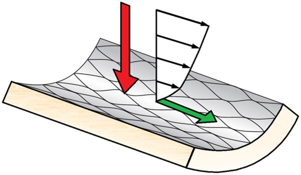
A schematic representation of the forces the vessel wall is exposed to. The red arrow is the normal force component, associated with blood pressure, and the green arrow is the tangential component force, associated with wall shear stress. Note that the size of the arrows does not represents the magnitude of the forces, only the direction.

A schematic illustration of some of the processes that are involved in plaque development and rupture, and how these are related to wall shear stress. In low wall shear stress regions, monocytes are recruited by the endothelium through the expression of various adhesion molecules. Recruited monocytes transform into foam cells which subsequently can accumulate and are the main source for the development of the necrotic core. Expansive remodelling initially prevents lumen intrusion of the plaque, but once this process fails, the wall shear patterns the plaque is exposed to changes. High wall shear stress will be present at the upstream part and the throat of the plaque, while in the downstream region, low wall shear stress will be accompanied by elevated levels of oscillatory flow. Although there is no direct effect of wall shear stress on plaque rupture, shear stress does influence endothelial function, potentially leading to cap thinning in the high wall shear stress regions.
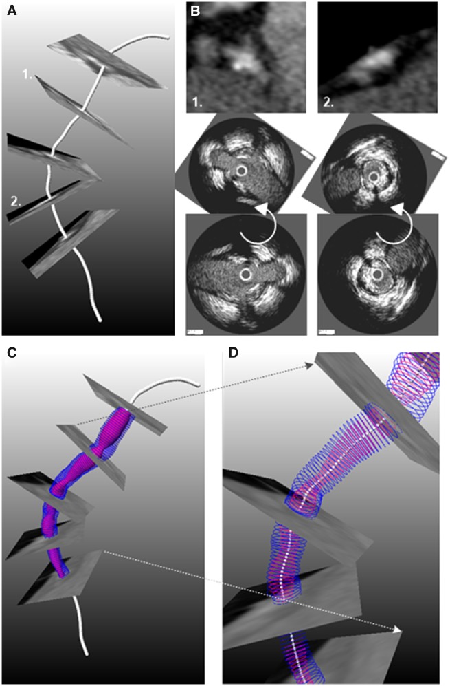
Methodology developed to fuse computed tomography angiography and intravascular imaging data: (A) Extraction of lumen centreline from computed tomography angiography. (B) Cross-sectional computed tomography angiography images are generated perpendicularly to the centreline and anatomical landmarks that are seen in intravascular ultrasound/optical coherence tomography images are detected. After matching the intravascular imaging contours are rotated so as to obtain the correct orientation (middle panel). (C) The orientated intravascular imaging contours are placed perpendicular to the lumen centreline extracted by computed tomography angiography to reconstruct the coronary artery anatomy that is shown in (D).
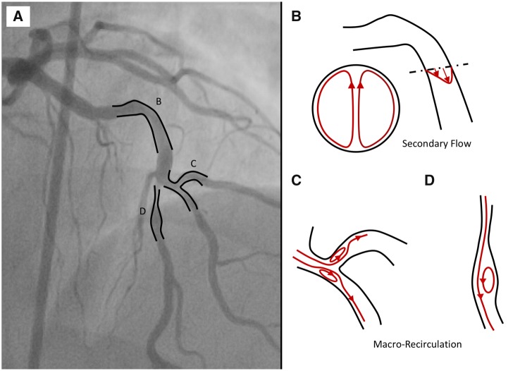
(A) Coronary tree showing multiple curvatures and bifurcation. (B) Secondary flow in the presence of the proximal curvature. (C) Macro-recirculation due to varying lumen geometries such as bifurcation and stenosis.
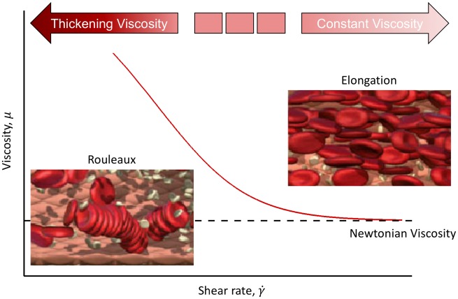
Changes in red blood cells under shear. Red blood cells aggregate at low shear rate, resulting in high blood viscosity. The process is reversed as shear rate increases.

Overview of micro flow environment around a stent strut (left) and the resulting high and low wall shear stress regions around the strut (right).
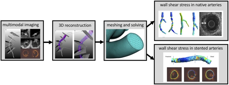
Wall shear stress in coronary arteries from imaging to modelling.
Similar articles
-
Wall shear stress: theoretical considerations and methods of measurement.Prog Cardiovasc Dis. 2007 Mar-Apr;49(5):307-29. doi: 10.1016/j.pcad.2006.11.001. Prog Cardiovasc Dis. 2007. PMID: 17329179 Review.
-
Assessment of superficial coronary vessel wall deformation and stress: validation of in silico models and human coronary arteries in vivo.Int J Cardiovasc Imaging. 2018 Jun;34(6):849-861. doi: 10.1007/s10554-018-1311-7. Epub 2018 Feb 3. Int J Cardiovasc Imaging. 2018. PMID: 29397475
-
Magnetohydrodynamic blood flow in patients with coronary artery disease.Comput Methods Programs Biomed. 2018 Sep;163:111-122. doi: 10.1016/j.cmpb.2018.06.007. Epub 2018 Jun 17. Comput Methods Programs Biomed. 2018. PMID: 30119846
-
Haemodynamic assessment of human coronary arteries is affected by degree of freedom of artery movement.Comput Methods Biomech Biomed Engin. 2017 Feb;20(3):260-272. doi: 10.1080/10255842.2016.1215439. Epub 2016 Jul 28. Comput Methods Biomech Biomed Engin. 2017. PMID: 27467730
-
A review study on blood in human coronary artery: Numerical approach.Comput Methods Programs Biomed. 2020 Apr;187:105243. doi: 10.1016/j.cmpb.2019.105243. Epub 2019 Nov 27. Comput Methods Programs Biomed. 2020. PMID: 31805457 Review.
Cited by
-
2'-5' oligoadenylate synthetase‑like 1 (OASL1) protects against atherosclerosis by maintaining endothelial nitric oxide synthase mRNA stability.Nat Commun. 2022 Nov 4;13(1):6647. doi: 10.1038/s41467-022-34433-z. Nat Commun. 2022. PMID: 36333342 Free PMC article.
-
A Synergistic Overview between Microfluidics and Numerical Research for Vascular Flow and Pathological Investigations.Sensors (Basel). 2024 Sep 10;24(18):5872. doi: 10.3390/s24185872. Sensors (Basel). 2024. PMID: 39338617 Free PMC article. Review.
-
Diabetic atherosclerosis: is there a role for the hypoxia-inducible factors?Biosci Rep. 2020 Aug 28;40(8):BSR20200026. doi: 10.1042/BSR20200026. Biosci Rep. 2020. PMID: 32816039 Free PMC article. Review.
-
Medical Image-Based Computational Fluid Dynamics and Fluid-Structure Interaction Analysis in Vascular Diseases.Front Bioeng Biotechnol. 2022 Apr 27;10:855791. doi: 10.3389/fbioe.2022.855791. eCollection 2022. Front Bioeng Biotechnol. 2022. PMID: 35573253 Free PMC article. Review.
-
Association Among Local Hemodynamic Parameters Derived From CT Angiography and Their Comparable Implications in Development of Acute Coronary Syndrome.Front Cardiovasc Med. 2021 Sep 13;8:713835. doi: 10.3389/fcvm.2021.713835. eCollection 2021. Front Cardiovasc Med. 2021. PMID: 34589527 Free PMC article.
References
-
- Young DF, Tsai FY.. Flow characteristics in models of arterial stenoses. I. Steady flow. J Biomech 1973;6:395–410. - PubMed
-
- Young DF, Tsai FY.. Flow characteristics in models of arterial stenoses. II. Unsteady flow. J Biomech 1973;6:547–559. - PubMed
-
- Gould KL, Kelley KO, Bolson EL.. Experimental validation of quantitative coronary arteriography for determining pressure-flow characteristics of coronary stenosis. Circulation 1982;66:930–937. - PubMed
-
- Kwak BR, Back M, Bochaton-Piallat ML, Caligiuri G, Daemen MJ, Davies PF, Hoefer IE, Holvoet P, Jo H, Krams R, Lehoux S, Monaco C, Steffens S, Virmani R, Weber C, Wentzel JJ, Evans PC.. Biomechanical factors in atherosclerosis: mechanisms and clinical implications. Eur Heart J 2014;35:3013–3020; 3020a–3020d. - PMC - PubMed
-
- Passerini AG, Polacek DC, Shi C, Francesco NM, Manduchi E, Grant GR, Pritchard WF, Powell S, Chang GY, Stoeckert CJ, Davies PF.. Coexisting proinflammatory and antioxidative endothelial transcription profiles in a disturbed flow region of the adult porcine aorta. Proc Natl Acad Sci USA 2004;101:2482–2487. - PMC - PubMed
Publication types
MeSH terms
Grants and funding
LinkOut - more resources
Full Text Sources
Other Literature Sources
Medical
