Review
. 2019 Oct;593(20):2925-2949.
doi: 10.1002/1873-3468.13627. Epub 2019 Oct 20.

Diverse roles for CDK-associated activity during spermatogenesis

Affiliations
Review

Diverse roles for CDK-associated activity during spermatogenesis

Nathan Palmer et al. FEBS Lett. 2019 Oct.

Abstract

The primary function of cyclin-dependent kinases (CDKs) in complex with their activating cyclin partners is to promote mitotic division in somatic cells. This canonical cell cycle-associated activity is also crucial for fertility as it allows the proliferation and differentiation of stem cells within the reproductive organs to generate meiotically competent cells. Intriguingly, several CDKs exhibit meiosis-specific functions and are essential for the completion of the two reductional meiotic divisions required to generate haploid gametes. These meiosis-specific functions are mediated by both known CDK/cyclin complexes and meiosis-specific CDK-regulators and are important for a variety of processes during meiotic prophase. The majority of meiotic defects observed upon deletion of these proteins occur during the extended prophase I of the first meiotic division. Importantly a lack of redundancy is seen within the meiotic arrest phenotypes described for many of these proteins, suggesting intricate layers of cell cycle control are required for normal meiotic progression. Using the process of male germ cell development (spermatogenesis) as a reference, this review seeks to highlight the diverse roles of selected CDKs their activators, and their regulators during gametogenesis.

Keywords: cyclin; cyclin-dependent kinase; meiosis; meiotic crossover; recombination; synapsis.


Figures

Figure 1
Figure 1
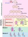
Schematic of adult spermatogenesis. Spermatogenesis is initiated by A single (As) type spermatogonial stem cells (SSCs). As type SSCs are thought to be able to produce both undifferentiated As type SSCs (self‐renewal) or separate populations of As SSCs capable of differentiation. As spermatogonia undergo consecutive rounds of mitotic division to produce pairs (Apr) or aligned chains (Aal) of SSCs. Aal SSCs represent clonal populations linked by shared cytoplasm and vary in the maximal chain length attained before further differentiation. Chains of 8 (Aal8) or more are considered proficient to enter further differentiation. Each Aal spermatogonia differentiates without division to form ‘differentiating’ A1 type spermatogonia. These undergo consecutive rounds of mitotic division to produce A2, A3, A3, Intermediate (In), and B‐type spermatogonia. B‐type spermatogonia subsequently differentiate without division to form primary spermatocytes capable of meiotic division. In premeiotic S phase, diploid primary spermatocytes duplicate their DNA to become tetraploid. During meiotic prophase I, chromosomal homologs initiate meiotic recombination and synapsis allowing the formation of meiotic crossovers. Upon resolution of crossovers, homologs are segregated into two diploid secondary spermatocytes (meiosis I). Each secondary spermatocyte divides rapidly to segregate sister chromatids into two haploid round spermatids (meiosis II). Thus, four haploid spermatids can be produced from two reductional divisions of a primary spermatocyte. Not shown here is the process of spermiogenesis whereby round spermatids undergo sequential steps of differentiation to form elongating spermatids and spermatozoa.
Figure 2
Figure 2
The roles of CDK2 and Speedy A in bouquet formation and synapsis. (1) During leptonema, Speedy A (red ovals) localizes to the inner nuclear envelope and interacts with the shelterin complex (yellow circles) on the telomeric ends of chromosomal homologs. This allows priming attachment or initial tethering of telomeres to the inner nuclear envelope. CDK2 (green circles) is then loaded onto telomeres in a manner dependent upon Speedy A. Here blue and pink lines are used to represent duplicated paternal and maternal chromosomal homologs, respectively; that is, each line contains two sister chromatids linked via their centromeres. (2) During zygonema, the interaction between shelterin, CDK2, and Speedy A is important in promoting the stable formation of the Linker of Nucleoskeleton and Cytoskeleton (LINC) complex. A simplified representation of the main components of the LINC complex is shown as a blow up below. Formation of a stable LINC complex is likely mediated by interactions between CDK2 and the inner nuclear envelope protein SUN1 (orange rectangles). SUN1 in turn is linked to the outer nuclear envelope by interactions with KASH5 (teal ovals). Cytoskeletal forces mediated by microtubules (gray) act on the LINC complex via KASH5 to drive telomere movement along the inner nuclear envelope. This allows telomeres to be polarized on the inner nuclear envelope leading to the formation of a characteristic bouquet pattern of chromosomes. (3) Bouquet formation in pachynema brings chromosomal homologs into close proximity and facilitates homolog paring (synapsis), which is completed in pachynema. An example of meiotic defect occurring upon impaired formation and/or stability of the LINC complex is shown at the bottom left hand side. These defects are characteristic of Cdk2−/− and Speedy A−/− spermatocytes and also mutants whereby shelterin or LINC complex components have been deleted.
Figure 3
Figure 3
Schematic of the crossover designation process. Upper box: pattern of early and late recombination nodule formation during meiotic prophase I. During early pachynema, proteins associated with early recombination nodules (early RN) localize as many distinct foci along the chromosomal axes. Here, paired chromosomal axes are represented by a red line and early recombination nodules are represented by blue circles. By mid‐pachynema, crossover designation leads to the selection of 1 or maximally 2 early recombination nodules to mature into late recombination nodules (late RN) as shown by green circles. At the same time, nondesignated early recombination nodules are repaired and their associated proteins dissociate from the chromosomal axes. By late pachynema, only late RNs remain associated with the chromosomal axes. These mark the sites at which meiotic crossovers will form. Lower box: Events leading to crossover designation or crossover de‐selection at early recombination nodules. During early pachynema, early RNs exhibit specific localization of many proteins including the MutSγ complex comprised of MSH4 (blue ring) and MSH5 (red ring). MutSγ is thought to be stabilized by SUMOylation mediated by RNF212 (yellow square). During mid‐pachynema, one of two possibilities can occur at early RNs, crossover designation (left hand side) or crossover de‐selection (right hand side). Crossover de‐selection occurs when RNF212 becomes destabilized and dissociates from early RNs. This is thought to allow the ubiquitin ligase CCNBIP1 (HEI10) (red square) to target early RN proteins such as MutSγ for ubiquitination and degradation. These events are associated with the downstream repair of early RNs as non‐crossovers. Crossover designation occurs when RNF212 remains stabilized at early RNs. This is associated with the localization of HEI10 and CDK2. These events are characteristic of late RN formation and leads to the downstream recruitment of the MutLγ complex comprised of MLH1 (green ring) and MLH3 (purple ring). These events are required for the downstream repair of late RNs as crossovers.
Figure 4
Figure 4
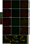
The pattern of CDK2 localization to chromosomal axes during meiotic prophase I. During leptonema, CDK2 foci (green) can be observed to localize to the telomeres of each chromosome. At this stage, chromosomal axes have not yet been formed as synaptonemal complex formation as determined by SYCP3 staining (red) is still at an early stage. During zygonema, homolog pairing is initiated and distinct axes of SYCP3 can be observed to associate with each other. At this stage, CDK2 localization is still specific to telomeres. During mid‐pachynema, intense singular CDK2 foci can be observed at paired telomeres of homologs. At this point, 1‐2 weaker but distinct interstitial foci of CDK2 can be observed marking the formation of late recombination nodules. In the lower most panel, several examples of paired homologs are shown at higher magnification to indicate both telomeric (T) and late recombination nodule‐associated (LRN) CDK2 foci. At this stage, staining can also be seen on the X‐Y chromosomes, but this is not addressed within the scope of this review. By diplonema, late recombination nodule‐associated CDK2 foci dissociate from the chromosomal axes but can still be observed as pairs of CDK2 foci at chromosomal ends. These represent the individual telomeres of each homolog, which separate upon the splitting of chromosomal axes. Scale bars for each image are shown in white and are equivalent to 5 µm in all pictures.

Similar articles

Cited by

References

    1. Morgan DO (1997) Cyclin‐dependent kinases: engines, clocks, and microprocessors. Annu Rev Cell Dev Biol 13, 261–291. - PubMed
    1. Cao L, Chen F, Yang X, Xu W, Xie J and Yu L (2014) Phylogenetic analysis of CDK and cyclin proteins in premetazoan lineages. BMC Evol Biol 14, 10. - PMC - PubMed
    1. Doonan JH and Kitsios G (2009) Functional evolution of cyclin‐dependent kinases. Mol Biotechnol 42, 14–29. - PubMed
    1. Lim S and Kaldis P (2013) Cdks, cyclins and CKIs: roles beyond cell cycle regulation. Development 140, 3079–3093. - PubMed
    1. Loyer P, Trembley JH, Katona R, Kidd VJ and Lahti JM (2005) Role of CDK/cyclin complexes in transcription and RNA splicing. Cell Signal 17, 1033–1051. - PubMed

Publication types

MeSH terms