

Management of gastric glomus tumor: A case report
- PMID: 31567933
- PMCID: PMC6756591
- DOI: 10.1097/MD.0000000000016980
Management of gastric glomus tumor: A case report
Abstract
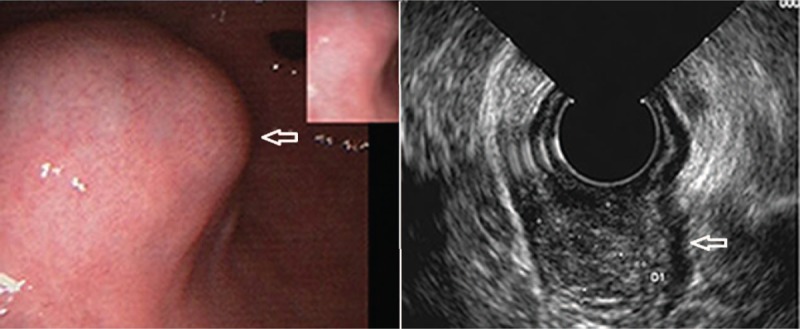
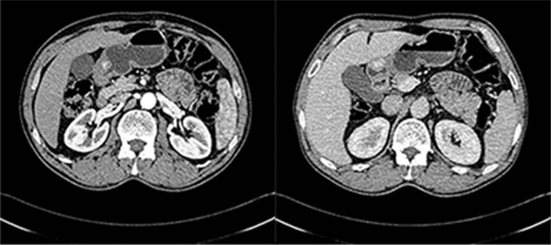
Rationale: Gastric glomus tumor (GGT) is a rare gastrointestinal tumor and its preoperative imaging features are significant to make a correct diagnosis, while the assessment of the pathological and immunohistochemical characteristics of the specimen are the main methods used for its diagnosis. This study introduces the clinical uniqueness, endoscopic ultrasonography, radiology, histology and immunohistochemistry results of a patient with GGT to discuss the imaging and clinico-pathological features, diagnosis and differential diagnosis of GGT.
Patient concerns: The patient expressed a complaint concerning an "intermittent abdominal pain for 4 months".
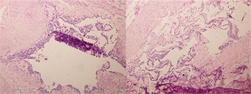
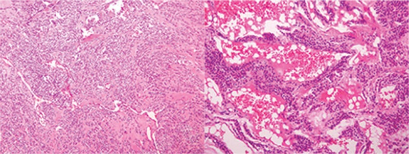
Diagnoses: The patient was diagnosed with gastric stromal tumor according to the clinical manifestations and imaging examination before the operation. The pathological examination of an intra-operative frozen sample confirmed the benign nature of the tumor, while post-operative immunohistochemistry results indicate the presence of a GGT. The postoperative histology revealed a tumor tissue composed of irregular blood vessels and glomus cells of same size with interstitial hyaline and mucoid degeneration. Immunohistochemical staining showed positivity for SMA (+), vimentin (3+), CD 34 (vascular +), and Factor VIII (vascular +).
Interventions: The tumor was completely removed by surgery.
Outcomes: The patient recovered well, and was discharged from the hospital. Five months after the operation, a normal gastric mucosa was observed by gastroscopic examination.
Lessons: Most of the GGTs are benign lesions, surgical resection is the preferred treatment and they result in a good prognosis. However, malignant GGT should be treated as soon as possible because of its metastatic potential and recurrence. Adjuvant radiotherapy or chemotherapy might be useful after operation.
Conflict of interest statement
The authors have no conflicts of interests to disclose.
Figures
References
-
- Hae-Wan Lee, Jae Jung Lee, Dae Hyun Yang, et al. A clinicopathologic study of glomus tumor of the stomach. J Clin Gastroenterol 2006;40:717–20. - PubMed
Publication types
MeSH terms
LinkOut - more resources
Full Text Sources
Medical
Miscellaneous
