. 2019 Dec;23(12):8010-8018.
doi: 10.1111/jcmm.14653. Epub 2019 Sep 30.

Non-lethal proteasome inhibition activates pro-tumorigenic pathways in multiple myeloma cells

Affiliations

Non-lethal proteasome inhibition activates pro-tumorigenic pathways in multiple myeloma cells

Aikaterini Skorda et al. J Cell Mol Med. 2019 Dec.

Abstract

Multiple myeloma (MM) is a haematological malignancy being characterized by clonal plasma cell proliferation in the bone marrow. Targeting the proteasome with specific inhibitors (PIs) has been proven a promising therapeutic strategy and PIs have been approved for the treatment of MM and mantle-cell lymphoma; yet, while outcome has improved, most patients inevitably relapse. As relapse refers to MM cells that survive therapy, we sought to identify the molecular responses induced in MM cells after non-lethal proteasome inhibition. By using bortezomib (BTZ), epoxomicin (EPOX; a carfilzomib-like PI) and three PIs, namely Rub999, PR671A and Rub1024 that target each of the three proteasome peptidases, we found that only BTZ and EPOX are toxic in MM cells at low concentrations. Phosphoproteomic profiling after treatment of MM cells with non-lethal (IC10 ) doses of the PIs revealed inhibitor- and cell type-specific readouts, being marked by the activation of tumorigenic STAT3 and STAT6. Consistently, cytokine/chemokine profiling revealed the increased secretion of immunosuppressive pro-tumorigenic cytokines (IL6 and IL8), along with the inhibition of potent T cell chemoattractant chemokines (CXCL10). These findings indicate that MM cells that survive treatment with therapeutic PIs shape a pro-tumorigenic immunosuppressive cellular and secretory bone marrow microenvironment that enables malignancy to relapse.

Keywords: cytokines; kinases; multiple myeloma; proteasome; proteasome inhibitors.


Conflict of interest statement

The authors declare no conflict of interest.

Figures
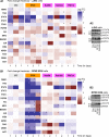
Figure 1
Figure 1
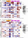
Non‐lethal inhibition of proteasome in MM cells activates pro‐survival, tumorigenic and immunosuppressive pathways in an inhibitor‐ and cell type‐specific manner. A1, Heatmap indicating logarithmic fold change (FC) values (vs control samples) of the basal phosphorylation levels of the shown proteins after incubating JJN3 cells with either BTZ and EPOX for 24, 48 and 168 h or with Rub999, Rub1024 and PR671A for 24 and 48 h. A2, Representative immunoblotting analyses of JJN3 cell protein samples probed with antibodies against p‐STAT3 and p‐STAT6 after exposure of cells to the shown PIs for 24 or 48 h. B1, Heatmap indicating logarithmic FC values (vs control samples) of the basal phosphorylation levels of the shown proteins in RPMI 8226 cells incubated with either BTZ and EPOX for 24, 48 and 168 h or with Rub999, Rub1024 and PR671A for 24 and 48 h. B2, Representative blots showing STAT3 and STAT6 phosphorylation levels after exposure of RPM1 8226 cells to the shown PIs for 24 or 48 h. Significance (*) of the results in (A1, B1) was set as a combination of median fluorescence intensity (MFI) value above 600 and FC value above 0.3 vs control samples. Probing with GAPDH in (A2, B2) was used as total protein loading reference
Figure 2
Figure 2
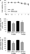
Combined proteasome and MTH1 inhibition exerted mild synergistic pro‐death effects on MM cells. A, Relative (%) viability of JJN3 and RPM1 8226 cell lines incubated with the MTH1 inhibitor TH588 for 24 h. B, C, Relative (%) survival of JJN3 (B) and RPM1 8226 (C) cell lines after a combinatorial treatment with BTZ or EPOX (at IC10 concentration) in the presence (or not) of the TH588 inhibitor for 24 h. BTZ (JJN3 cells, 2.45 nM; RPMI 8226, 1.8 nM), EPOX (JJN3 cells, 4.54 nM; RPMI 8226, 5.5 nM), TH588 (JJN3 cells, 5.5 μM; RPMI 8226, 9 μM). Bars: ± SD, *P < .05, **P < .01 vs controls set to 100%
Figure 3
Figure 3
Incubation of MM cell lines with non‐lethal doses of PIs promotes (in an inhibitor‐ and cell type‐specific manner) the secretion of pro‐tumorigenic immunosuppressive molecules. A1, Heatmap indicating logarithmic FC values (vs control samples) of the assayed cytokine/chemokine secretion levels after treatment of JJN3 cells with either BTZ and EPOX for 24, 48 and 168 h or with Rub999, PR671A and Rub1024 for 24 and 48 h. A2, Representative blots of JJN3 cell culture supernatant probed with antibodies against IL6, IL8 and CXCL10 after treatment with the shown PIs for 24 or 48 h. B1, Heatmap indicating logarithmic FC values (vs control samples) of the assayed cytokine/chemokine secretion levels in RPMI 8226 cells treated with either BTZ and EPOX for 24, 48 and 168 h or with Rub999, PR671A and Rub1024 for 24 and 48 h. B2, Representative blots showing the IL6, IL8 and CXCL10 levels in RPMI 8226 cell culture supernatant after exposure to the shown PIs for 24 or 48 h. Significance (*) of the results in (A1, B1) was set as a combination of median fluorescence intensity (MFI) value above 600 and fold change (FC) value above 0.3 vs control samples. Ponceau S staining of nitrocellulose membranes in (A2, B2) was used as reference for total protein input

References

    1. Trougakos IP, Sesti F, Tsakiri E, Gorgoulis VG. Non‐enzymatic post‐translational protein modifications and proteostasis network deregulation in carcinogenesis. J Proteomics. 2013;92:274‐298. - PubMed
    1. Sala AJ, Bott LC, Morimoto RI. Shaping proteostasis at the cellular, tissue, and organismal level. J Cell Biol. 2017;216:1231‐1241. - PMC - PubMed
    1. Sykiotis GP, Bohmann D. Stress‐activated cap'n'collar transcription factors in aging and human disease. Sci Signal. 2010;3:re3. - PMC - PubMed
    1. Tsakiri EN, Trougakos IP. The amazing ubiquitin‐proteasome system: structural components and implication in aging. Int Rev Cell Mol Biol. 2015;314:171‐237. - PubMed
    1. Vilchez D, Saez I, Dillin A. The role of protein clearance mechanisms in organismal ageing and age‐related diseases. Nat Commun. 2014;5:5659. - PubMed

Publication types

MeSH terms