. 2019 Sep 27;11(10):1446.
doi: 10.3390/cancers11101446.

In Vitro Characterization of Cisplatin and Pemetrexed Effects in Malignant Pleural Mesothelioma 3D Culture Phenotypes

Affiliations

In Vitro Characterization of Cisplatin and Pemetrexed Effects in Malignant Pleural Mesothelioma 3D Culture Phenotypes

Eleftherios D Papazoglou et al. Cancers (Basel). .

Abstract

Malignant pleural mesothelioma (MPM) is an aggressive cancer with poor prognosis. The main treatment for MPM is doublet chemotherapy with Cisplatin and Pemetrexed, while ongoing trials test the efficacy of pemetrexed monotherapy. However, there is lack of evidence regarding the effects of Cisplatin and Pemetrexed on MPM cell phenotypes, especially in three-dimensional (3D) cell cultures. In this study, we evaluated the effects Cisplatin and Pemetrexed on cell viability using homologous cell derived extracellular matrix (hECM) as substratum and subsequently in the following 3D cell culture phenotypes: tumor spheroid formation, tumor spheroid invasion, and collagen gel contraction. We used benign mesothelial MeT-5A cells as controls and the MPM cell lines M14K (epithelioid), MSTO (biphasic), and ZL34 (sarcomatoid). Cell viability of all cell lines was significantly decreased with all treatments. Mean tumor spheroid perimeter was reduced after treatment with Pemetrexed or the doublet therapy in all cell lines, while Cisplatin reduced the mean spheroid perimeter of MeT-5A and MSTO cells. Doublet treatment reduced the invasive capacity of spheroids of cell lines into collagenous matrices, while Cisplatin lowered the invasion of the MSTO and ZL34 cell lines, and Pemetrexed lowered the invasion of MeT-5A and ZL34 cell lines. Treatment with Pemetrexed or the combination significantly reduced the collagen gel contraction of all cell lines, while Cisplatin treatment affected only the MeT-5A and M14K cells. The results of the current study can be used as an in vitro 3D platform for testing novel drugs against MPM for ameliorating the effects of first line chemotherapeutics.

Keywords: 3D cultures; cisplatin; homologous cell derived extracellular matrix; malignant pleural mesothelioma; mesothelial cells; pemetrexed; pleura; tumor spheroid.


Conflict of interest statement

The authors declare no conflict of interest.

Figures

Figure 1
Figure 1
Mean values of cell viability ± SEM without or with treatments (CPDD, Pem, or CPDD+Pem) expressed as % of Controls (cells with 10% FBS-RPMI). (A) MeT-5A cells (n = 13), (B) M14K cells (n = 16), (C) MSTO cells (n = 16), (D) ZL34 (n = 15 or 16). *** p < 0.001 vs. Controls, @@ p < 0.01 and @@@ p < 0.001 vs. CPDD, # p < 0.05 vs. Pem.
Figure 2
Figure 2
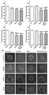
Mean values of spheroid perimeter ± SEM without or with treatments (CPDD, Pem, or CPDD+Pem) expressed as % of Controls (cells with 10% FBS-RPMI), along with representative microscopy images from each group. (A) MeT-5A cells (n = 13–17), (B) M14K cells (n = 10–15), (C) MSTO cells (n = 15–18), (D) ZL34 (n = 13–15). *** p < 0.001 vs. Controls, @@@ p < 0.001 vs. CPDD. (E) Images of spheroids of cell types arranged by rows and their corresponding treatments in a column wise manner. Scale bars represent a length of 100 pixels.
Figure 3
Figure 3
Mean values of perimeter of spheroid invasion ± SEM without or with treatments (CPDD, Pem, or CPDD+Pem) expressed as % of Controls (cells with 10% FBS-RPMI) along with representative microscopy images from each group. (A) MeT-5A cells (n = 16–21), (B) M14K cells (n = 19–22), (C) MSTO cells (n = 13–19), (D) ZL34 (n = 15–23). ** p < 0.01 and *** p < 0.001 vs. Controls, ## p < 0.01 vs. Pem. (E) Images of spheroids invading into the surrounding collagen matrix, arranged by rows (by cell type) and their corresponding treatments in a column wise manner. Scale bars represent a length of 100 pixels.
Figure 4
Figure 4
Mean values of gel area ± SEM without or with treatments (CPDD, Pem, or CPDD+Pem) expressed as % of Controls (cells with 10% FBS-RPMI) along with representative microscopy images from each group. (A) MeT-5A cells (n = 12), (B) M14K cells (n = 12), (C) MSTO cells (n = 16), (D) ZL34 (n = 11–12). ** p < 0.01 and *** p < 0.001 vs. Controls, @@ p < 0.01 and @@@ p < 0.001 vs. CPPD, ## p < 0.01 vs. Pem. (E) Images of contracted gels with cell types arranged by rows and their corresponding treatments in a column wise manner.

References

    1. Rossini M., Rizzo P., Bononi I., Clementz A., Ferrari R., Martini F., Tognon M.G. New Perspectives on Diagnosis and Therapy of Malignant Pleural Mesothelioma. Front. Oncol. 2018;8:91. doi: 10.3389/fonc.2018.00091. - DOI - PMC - PubMed
    1. Baumann F., Ambrosi J.P., Carbone M. Asbestos is not just asbestos: an unrecognised health hazard. Lancet Oncol. 2013;14:576–578. doi: 10.1016/S1470-2045(13)70257-2. - DOI - PubMed
    1. Bakker E., Guazzelli A., Krstic-Demonacos M., Lisanti M., Sotgia F., Mutti L. Current and prospective pharmacotherapies for the treatment of pleural mesothelioma. Expert Opin. Orphan Drugs. 2017;5:455–465. doi: 10.1080/21678707.2017.1325358. - DOI
    1. Baas P., Fennell D., Kerr K.M., Van Schil P.E., Haas R.L., Peters S. Malignant pleural mesothelioma: ESMO Clinical Practice Guidelines for diagnosis, treatment and follow-up. Ann. Oncol. 2015;26:31–39. doi: 10.1093/annonc/mdv199. - DOI - PubMed
    1. Nowak A.K. Chemotherapy for malignant pleural mesothelioma: a review of current management and a look to the future. Ann. Cardiothorac. Surg. 2012;1:508–515. - PMC - PubMed

LinkOut - more resources