Review
. 2019 Sep 27;11(10):1451.
doi: 10.3390/cancers11101451.

Adipocytes and microRNAs Crosstalk: A Key Tile in the Mosaic of Breast Cancer Microenvironment

Affiliations
Review

Adipocytes and microRNAs Crosstalk: A Key Tile in the Mosaic of Breast Cancer Microenvironment

Erika Bandini et al. Cancers (Basel). .

Abstract

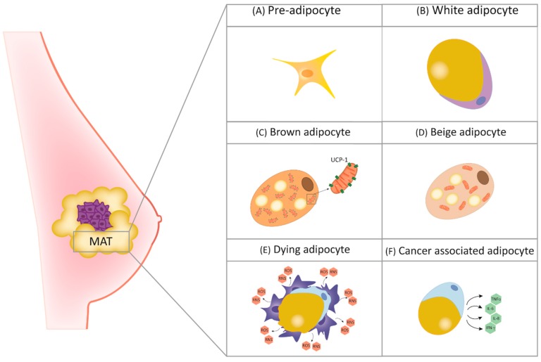
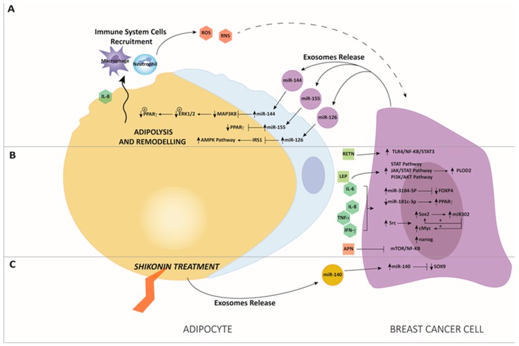
Breast cancer (BC) is a disease characterized by a high grade of heterogeneity. Consequently, despite the great achievements obtained in the last decades, most of the current therapeutic regimens still fail. The identification of new molecular mechanisms that will increase the knowledge of all steps of tumor initiation and growth is mandatory in finding new clinical strategies. The BC microenvironment, consisting of endothelial cells, fibroblasts, immune cells and adipocytes, plays an essential role in regulating BC development, and recently it has gained great attention in the scientific community. In particular, adipose tissue is emerging as an important target to investigate among mammary gland components. The mechanisms underlying BC progression driven by adipocytes are predominantly unexplored, especially that involving the switch from normal adipocytes to the so-called cancer-associated adipocytes (CAAs). MicroRNAs (miRNAs), a class of gene expression modulators, have emerged as the regulators of key oncogenes and tumor suppressor genes that affect multiple pathways of the tumor microenvironment and adipose tissue. This review concerns a presentation of the role of adipocytes in breast tissue, and describes the most recent discoveries about the interplay between adipocytes and miRNAs, which collaborate in the arrangement of a pro-inflammatory and cancerous microenvironment, laying the foundations for new concepts in the prevention and treatment of BC.

Keywords: Breast Cancer; adipocytes; microRNAs.


Conflict of interest statement

The authors declare no conflict of interest.

Figures

Figure 1
Figure 1
Typical mammary adipose tissue (MAT) cellular composition. (A) Pre-adipocytes, immature precursor of mature adipocytes; (B) white adipocytes, with lipid-storage and endocrine functions, contain single lipid droplet occupies the largest part of the cytoplasm; (C) brown adipocytes, containing several cytoplasmic small fatty droplets and mitochondria, with uncoupling protein 1 (UCP1)-dependent thermoregulatory functions; (D) beige adipocytes, with UCP1-independent thermoregulatory functions; (E) dying adipocytes, form crown-like structures due to macrophage recruitment, causing the release of reactive nitrogen species (RNS) and reactive oxygen species (ROS); (F) cancer-associated adipocytes, with unbalanced production of pro-inflammatory cytokines.
Figure 2
Figure 2
(A) After co-culture between adipocytes and breast cancer (BC) cells, high levels of miR-144 and miR-126 were released by exosomes by BC cells. This miRs delivery leads to final adipolysis and remodeling of adipose tissue driven by miR-144 through the downregulation of Mitogen-Activated Protein Kinase Kinase Kinase 8/Mitogen-Activated Protein Kinase 3/1/Peroxisome Proliferator Activated Receptor Gamma (MAPK3/ERK1/2/ pPPARγ) and by miR-126 through the Protein Kinase AMP-Activated Catalytic Subunit Alpha 1 (AMPK)/ autophagy pathway, causing adipocyte-induced tumor growth [77]. BC-derived exosomes after adipocytes interaction carry a higher level of miR-155, which promotes adipocytes differentiation and remodeling in surrounding adipocytes targeting pPPARγ, thus facilitating tumor progression [76]. Increased adipocyte-derived interleukin-8 (IL-8) is responsible for neutrophils and macrophages recruitment, leading to the production of Reactive Oxygen Species (ROS) and Reactive Nitrogen Species (RNS), which act as a threat to tumor initiation. (B) Interaction of adipocytes with BC cells increase the release of proinflammatory cytokines and chemokines, leading to an upregulation of miR-3184-5p and downregulation of miR-181c-3p in BC cells, with the modulation of their respective target genes, forkhead box P4 (FOXP4) and PPARα; increased release of adipocyte-derived proinflammatory cytokines and adipokins such as leptin (LEP), IL-6, IL-8 and tumor necrosis factor alpha (TNFα) is responsible for the induction of janus kinase/signal transducers and activators of transcription (JAK/STAT) and through phosphoinositide 3-kinase/protein kinase B (PIK3/AKT) pathways. Resistin (RETN) promotes BC progression through the Toll like receptor 4/nuclear factor kappa-light-chain-enhancer of activated B cells/ signal transducer and activator of transcription 3 (TLR4/ NF-κB /STAT3) axis. As a result, EMT, migration and invasion pathways are enhanced in BC cells [72]. Likewise, after adipocytes and BC cells co-culture, there is an increase of proinflammatory cytokines secretion, with an activation of Src that upregulates SRY-Box transcription factor 2 (SOX2), cMYC and Nanog. SOX2 induces miR-302b transcription that, in turn, further stimulates cMYC and SOX2 expression, increasing the cytokine-induced cancer stem cell-like properties [73]. Conversely, adiponectin (APN) has anti-tumoral properties by inhibiting mammalian target of rapamycin (mTOR) and NF-κB pathways. (C) Treatment with the anti-tumor compound shikonin (SK). SK-treated adipocytes release high levels of miR-140, which block tumor progression of co-cultured BC cells targeting SOX9 signaling [74].

Similar articles

Cited by

References

    1. Siegel R.L., Miller K.D., Jemal A. Cancer statistics, 2017. CA Cancer J. Clin. 2017;67:7–30. doi: 10.3322/caac.21387. - DOI - PubMed
    1. Zardavas D., Irrthum A., Swanton C., Piccart M. Clinical management of breast cancer heterogeneity. Nat. Rev. Clin. Oncol. 2015;12:381–394. doi: 10.1038/nrclinonc.2015.73. - DOI - PubMed
    1. Mao Y., Keller E.T., Garfield D.H., Shen K., Wang J. Stromal cells in tumor microenvironment and breast cancer. Cancer Metastasis Rev. 2013;32:303–315. doi: 10.1007/s10555-012-9415-3. - DOI - PMC - PubMed
    1. Vivacqua A., Muoio M., Miglietta A., Maggiolini M. Differential MicroRNA Landscape Triggered by Estrogens in Cancer Associated Fibroblasts (CAFs) of Primary and Metastatic Breast Tumors. Cancers (Basel) 2019;11:412. doi: 10.3390/cancers11030412. - DOI - PMC - PubMed
    1. Vu L.T., Peng B., Zhang D.X., Ma V., Mathey-Andrews C.A., Lam C.K., Kiomourtzis T., Jin J., McReynolds L., Huang L., et al. Tumor-secreted extracellular vesicles promote the activation of cancer-associated fibroblasts via the transfer of microRNA-125b. J. Extracell. Vesicles. 2019;8:1599680. doi: 10.1080/20013078.2019.1599680. - DOI - PMC - PubMed

LinkOut - more resources