The effect of needle tip position on the analgesic efficacy of pulsed radiofrequency treatment in patients with chronic lumbar radicular pain: a retrospective observational study
- PMID: 31569920
- PMCID: PMC6813899
- DOI: 10.3344/kjp.2019.32.4.280
The effect of needle tip position on the analgesic efficacy of pulsed radiofrequency treatment in patients with chronic lumbar radicular pain: a retrospective observational study
Abstract
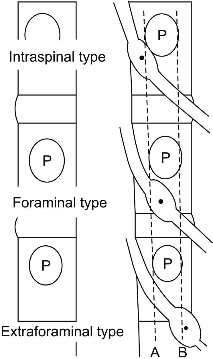
Background: Pulsed radiofrequency (PRF) is a treatment modality that alleviates radicular pain by intermittently applying high-frequency currents adjacent to the dorsal root ganglion. There has been no comparative study on analgesic effect according to the position of the needle tip in PRF treatment. The objective of this study is to evaluate the clinical outcomes of PRF according to the needle tip position.
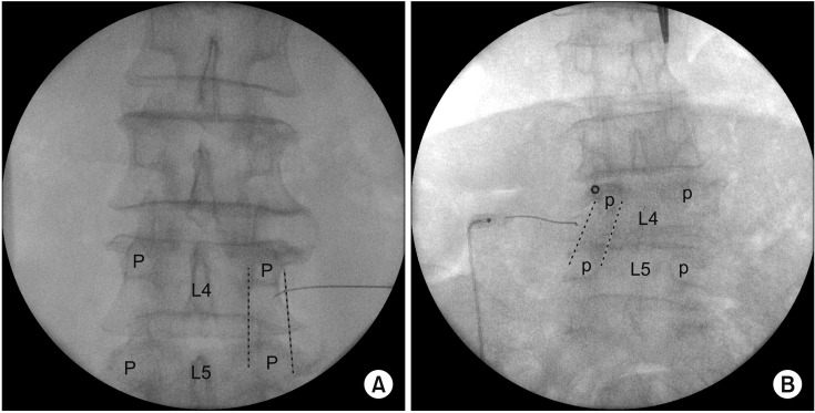
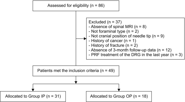
Methods: Patients were classified into 2 groups (group IP [group inside of pedicle] and group OP [group outside of pedicle]) based on needle tip position in the anteroposterior view of fluoroscopy. In the anteroposterior view, the needle tip was advanced medially further than the lateral aspect of the corresponding pedicle in group IP; however, in group OP, the needle tip was not advanced. The treatment outcomes and pain scores were evaluated at 4, 8, and 12 weeks after applying PRF.
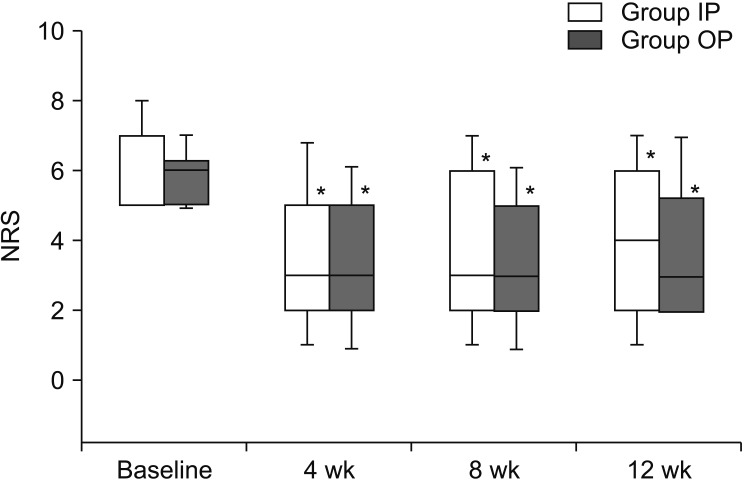
Results: At 4, 8, and 12 weeks, there were no significant differences between the successful response rate and numerical rating scale score ratio.
Conclusions: The analgesic efficacy of PRF treatment did not differ with the needle tip position.
Keywords: Analgesics; Ganglia, Spinal; Low Back Pain; Lumbosacral Region; Needles; Pulsed Radiofrequency Treatment; Radiculopathy; Spinal Nerve Roots.
Conflict of interest statement
No potential conflict of interest relevant to this article was reported.
Figures
Similar articles
-
Comparison of two distinct needle tip positions in pulsed radiofrequency for herpes zoster-related pain.CNS Neurosci Ther. 2023 Jul;29(7):1881-1888. doi: 10.1111/cns.14146. Epub 2023 Mar 7. CNS Neurosci Ther. 2023. PMID: 36880287 Free PMC article. Clinical Trial.
-
The Effect of Bipolar Pulsed Radiofrequency Treatment on Chronic Lumbosacral Radicular Pain Refractory to Monopolar Pulsed Radiofrequency Treatment.Pain Physician. 2018 Mar;21(2):E97-E103. Pain Physician. 2018. PMID: 29565952
-
Output Current and Efficacy of Pulsed Radiofrequency of the Lumbar Dorsal Root Ganglion in Patients With Lumbar Radiculopathy: A Prospective, Double-blind, Randomized Pilot Study.Pain Physician. 2023 Nov;26(7):E797-E804. Pain Physician. 2023. PMID: 37976483 Clinical Trial.
-
Pulsed radiofrequency treatment in interventional pain management: mechanisms and potential indications-a review.Acta Neurochir (Wien). 2011 Apr;153(4):763-71. doi: 10.1007/s00701-010-0881-5. Epub 2010 Nov 30. Acta Neurochir (Wien). 2011. PMID: 21116663 Free PMC article. Review.
-
A comprehensive review of pulsed radiofrequency in the treatment of pain associated with different spinal conditions.Br J Radiol. 2017 May;90(1073):20150406. doi: 10.1259/bjr.20150406. Epub 2017 Feb 10. Br J Radiol. 2017. PMID: 28186832 Free PMC article. Review.
Cited by
-
Comparison of two distinct needle tip positions in pulsed radiofrequency for herpes zoster-related pain.CNS Neurosci Ther. 2023 Jul;29(7):1881-1888. doi: 10.1111/cns.14146. Epub 2023 Mar 7. CNS Neurosci Ther. 2023. PMID: 36880287 Free PMC article. Clinical Trial.
-
The treatment of persistent spinal pain syndrome with epidural pulsed radiofrequency: improvement of the technique.Front Neurol. 2023 Oct 16;14:1236270. doi: 10.3389/fneur.2023.1236270. eCollection 2023. Front Neurol. 2023. PMID: 37909029 Free PMC article.
-
Observation and measurement of applied anatomical features for thoracic intervertebral foramen puncture on computed tomography images.World J Clin Cases. 2021 Jun 26;9(18):4607-4616. doi: 10.12998/wjcc.v9.i18.4607. World J Clin Cases. 2021. PMID: 34222427 Free PMC article.
-
A Literature Review of Dorsal Root Entry Zone Complex (DREZC) Lesions: Integration of Translational Data for an Evolution to More Accurate Nomenclature.J Pain Res. 2021 Jan 7;14:1-12. doi: 10.2147/JPR.S255726. eCollection 2021. J Pain Res. 2021. PMID: 33442287 Free PMC article. Review.
-
Optimal cutoff point of vertebral body cross-sectional area as a morphological parameter for predicting lumbar spondylolysis.Medicine (Baltimore). 2023 Sep 15;102(37):e35173. doi: 10.1097/MD.0000000000035173. Medicine (Baltimore). 2023. PMID: 37713872 Free PMC article.
References
-
- Sugawara O, Atsuta Y, Iwahara T, Muramoto T, Watakabe M, Takemitsu Y. The effects of mechanical compression and hypoxia on nerve root and dorsal root ganglia. an analysis of ectopic firing using an in vitro model. Spine (Phila Pa 1976) 1996;21:2089–94. doi: 10.1097/00007632-199609150-00006. - DOI - PubMed
LinkOut - more resources
Full Text Sources
Medical
Miscellaneous