

Ultra-High-Resolution Imaging of Amygdala Subnuclei Structural Connectivity in Major Depressive Disorder
- PMID: 31570286
- PMCID: PMC7010542
- DOI: 10.1016/j.bpsc.2019.07.010
Ultra-High-Resolution Imaging of Amygdala Subnuclei Structural Connectivity in Major Depressive Disorder
Abstract
Background: Major depressive disorder (MDD) is an increasingly common and disabling illness. As the amygdala has been reported to have pathological involvement in mood disorders, we aimed to investigate for the first time potential changes to structural connectivity of individual amygdala subnuclei in MDD using ultra-high-field 7T diffusion magnetic resonance imaging.
Methods: Twenty-four patients with MDD (11 women) and 24 age-matched healthy control participants (7 women) underwent diffusion-weighted imaging with a 1.05-mm isotropic resolution at 7T. Amygdala nuclei regions of interest were obtained through automated segmentation of 0.69-mm resolution T1-weighted images and 0.35-mm resolution T2-weighted images. Probabilistic tractography was performed on all subjects, with random seeding at each amygdala nucleus.
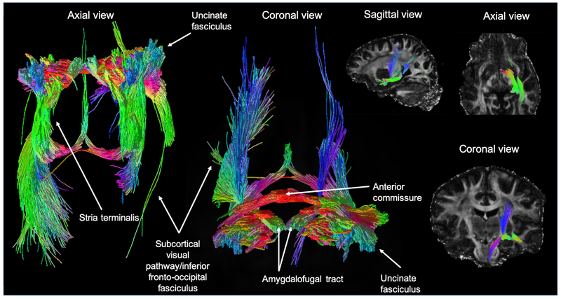
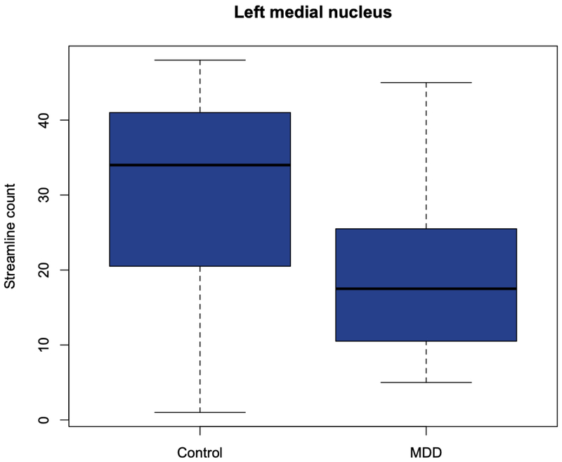
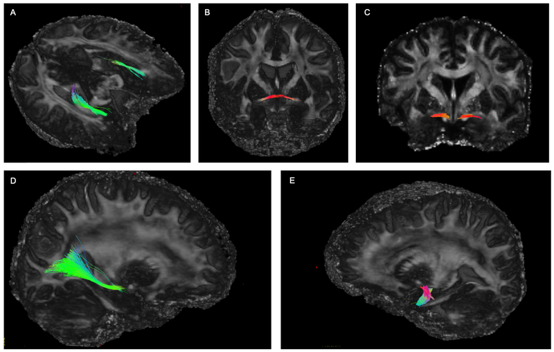
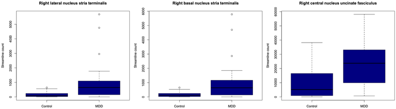
Results: The right lateral, basal, central, and centrocortical amygdala nuclei exhibited significantly increased connection density to the rest of the brain, whereas the left medial nucleus demonstrated significantly lower connection density (false discovery rate p < .05). Increased connection density in the right lateral and basal nuclei was driven by the stria terminalis, and the significant difference in the right central nucleus was driven by the uncinate fasciculus. Decreased connection density at the left medial nucleus did not appear to be driven by any individual white matter tract.
Conclusions: By exploiting ultra-high-resolution magnetic resonance imaging, structural hyperconnectivity was demonstrated involving the amygdaloid nuclei in the right hemisphere in MDD. To a lesser extent, impairment of subnuclei connectivity was shown in the left hemisphere.
Keywords: 7T; Amygdala; Diffusion MRI; Major depressive disorder; Neuroimaging; Tractography; Ultra-high-field.
Copyright © 2019 Society of Biological Psychiatry. Published by Elsevier Inc. All rights reserved.
Conflict of interest statement
Conflict of interest
Dr. Balchandani (the Principal Investigator in this study) is a named inventor on patents relating to magnetic resonance imaging (MRI) and RF pulse design. The patents have been licensed to GE Healthcare, Siemens AG, and Philips international. Dr. Balchandani receives royalty payments relating to these patents.
In the past 5 years, Dr. Murrough has provided consultation services to Boehreinger Ingelheim, Sage Therapeutics, FSV7, Novartis, Allergan, Fortress Biotech, Janssen Research and Development, Medavante-Prophase and Global Medical Education (GME) and has received research support from Avanir Pharmaceuticals, Inc.
No other authors reported biomedical financial interests or potential conflicts of interest.
Figures

References
-
- Conwell Y, Duberstein PR, Cox C, Herrmann JH, Forbes NT, Caine ED (1996): Relationships of age and axis I diagnoses in victims of completed suicide: a psychological autopsy study. Am J Psychiatry. 153:1001–1008. - PubMed
-
- Cuijpers P, Smit F (2002): Excess mortality in depression: a meta-analysis of community studies. J Affect Disord. 72:227–236. - PubMed
-
- Tang Y, Wang F, Xie G, Liu J, Li L, Su L, et al. (2007): Reduced ventral anterior cingulate and amygdala volumes in medication-naive females with major depressive disorder: A voxel-based morphometric magnetic resonance imaging study. Psychiatry Res. 156:83–86. - PubMed
-
- Bora E, Fornito A, Pantelis C, Yucel M (2012): Gray matter abnormalities in Major Depressive Disorder: a meta-analysis of voxel based morphometry studies. J Affect Disord. 138:9–18. - PubMed
Publication types
MeSH terms
Grants and funding
LinkOut - more resources
Full Text Sources
Research Materials
