Review
. 2020 May 1;12(5):a035055.
doi: 10.1101/cshperspect.a035055.

Store-Operated Calcium Channels: From Function to Structure and Back Again

Affiliations
Review

Store-Operated Calcium Channels: From Function to Structure and Back Again

Richard S Lewis. Cold Spring Harb Perspect Biol. .

Abstract

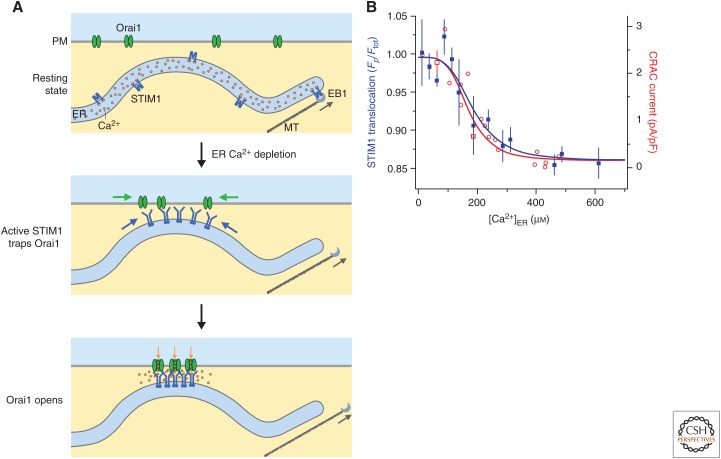
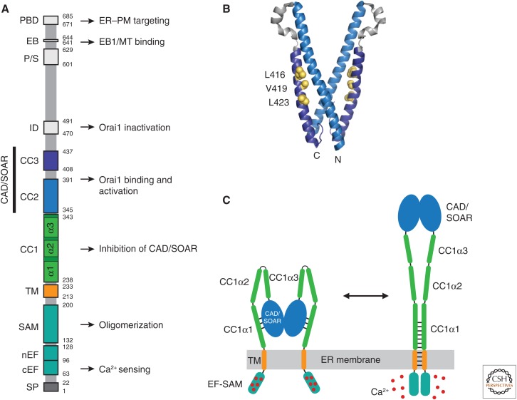
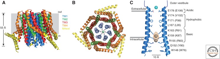
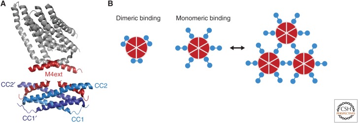
Store-operated calcium (Ca2+) entry (SOCE) occurs through a widely distributed family of ion channels activated by the loss of Ca2+ from the endoplasmic reticulum (ER). The best understood of these is the Ca2+ release-activated Ca2+ (CRAC) channel, which is notable for its unique activation mechanism as well as its many essential physiological functions and the diverse pathologies that result from dysregulation. In response to ER Ca2+ depletion, CRAC channels are formed through a diffusion trap mechanism at ER-plasma membrane (PM) junctions, where the ER Ca2+-sensing stromal interaction molecule (STIM) proteins bind and activate hexamers of Orai pore-forming proteins to trigger Ca2+ entry. Cell biological studies are clarifying the architecture of ER-PM junctions, their roles in Ca2+ and lipid transport, and functional interactions with cytoskeletal proteins. Molecular structures of STIM and Orai have inspired a multitude of mutagenesis and electrophysiological studies that reveal potential mechanisms for how STIM is toggled between inactive and active states, how it binds and activates Orai, and the importance of STIM-binding stoichiometry for opening the channel and establishing its signature characteristics of extremely high Ca2+ selectivity and low Ca2+ conductance.


Figures

Figure 1.
Figure 1.
Cellular choreography of store-operated calcium channels. (A) (Top) In resting cells with full endoplasmic reticulum (ER) Ca2+ stores, STIM1 and Orai1 diffuse freely in the ER and plasma membrane (PM), respectively. STIM1 also binds to EB1 at the tips of growing microtubules (MTs), which extends ER tubules toward the cell periphery. (Middle) After store depletion, STIM1 is activated, detaches from EB1, and accumulates at ER–PM junctions through interactions with negatively charged inositol phospholipids in the PM. Orai1 diffusing in the PM is trapped through binding to the CAD/SOAR domain of STIM1. (Bottom) Binding of STIM1 to the six Orai1 subunits of the Ca2+ release-activated Ca2+ (CRAC) channel opens the channel to allow local Ca2+ entry. (B) STIM1 translocation to ER–PM junctions (blue, measured by the ratio of labeled STIM1 at the periphery to the total STIM1 in the cell, Fp/Ftot) and CRAC channel current density (red) show a similar steep dependence on ER Ca2+ concentration ([Ca2+]ER). (Panel B is from Luik et al. 2008; adapted, with permission, from HHS Public Access and Nature Publishing.)
Figure 2.
Figure 2.
STIM1 functional domains and a model for activation. (A) Domain organization of STIM1. Functional domains include the signal peptide (SP), canonical EF-hand (cEF), noncanonical EF-hand (nEF), sterile α motif (SAM), transmembrane (TM) domain, putative coiled-coil domains 1–3 (CC1–3), Ca2+ release-activated Ca2+ (CRAC) activation domain (CAD, aa 342–448) or STIM-Orai-activating region (SOAR, aa 344–442), inactivation domain (ID), proline/serine-rich domain (P/S), EB1-binding (EB) domain, and the polybasic domain (PBD). Known functions are indicated. (B) Crystal structure of the dimeric CAD/SOAR (3TEQ.pdb) (Yang et al. 2012). Amino- and carboxy-terminal ends of the front subunit and selected residues implicated in interactions with CC1 (L416, V419, L423) are indicated in yellow. (C) A coiled-coil model for STIM1 activation by ER Ca2+ depletion (regions downstream from CAD/SOAR omitted for clarity). In the resting state (left), the luminal EF-SAM domains are bound to 5–6 Ca2+ ions and separated, which allows CC1α1 to bind to CAD/SOAR and sequester it close to the endoplasmic reticulum (ER) membrane. Following store depletion (right), Ca2+ release triggers dimerization of the EF-SAM domain, bringing the TM domains together to form a coiled-coil. This rearrangement favors dissociation of CC1α1 from CAD/SOAR and extends the coiled-coil beyond the ER membrane to move CAD/SOAR toward the plasma membrane (PM).
Figure 3.
Figure 3.
The closed-channel structure of Drosophila Orai (dOrai). (A) Side view of the crystal structure of dOrai, showing the arrangement of the transmembrane (TM) domains of the subunits (4HKR.pdb). (Panel A created based on data in Hou et al. 2012.) (B) Top view of dOrai, showing the six TM1 subunits arranged around a central pore, an interlocking cage of TM2/TM3 subunits, and peripheral TM4 subunits with three paired coiled-coil interactions of the cytoplasmic M4ext helices. (C) Side view of TM1 and amino-terminal extension helices from two opposed subunits in the crystal structure, showing pore-lining residues and functional pore domains. The corresponding human Orai1 residues are shown in parentheses. A Ba2+ ion is shown in blue above E178. The anion density with Fe atoms modeled into the structure are shown in yellow and gray in the inner pore.
Figure 4.
Figure 4.
Dimeric and monomeric models of STIM1-Orai1 binding. (A) A dimeric binding model, showing two subunits from the crystal structure of dOrai (top), with the cytoplasmic M4ext regions highlighted in red (4HKR.pdb) (Hou et al. 2012). The nuclear magnetic resonance (NMR) spectroscopy structure below shows two corresponding human Orai1 M4ext fragments (aa 272–292) bound to a dimer of CC1–CC2 STIM1 fragments (aa 312–383; 2MAK.pdb) (Stathopulos et al. 2013). (B) Schematic views of dimeric and monomeric binding models in which three or six STIM1 dimers (blue) are bound to the six Orai1 subunits (red), respectively. Monomeric binding may allow STIM1 dimers to create cross-linked arrays of channels (right).
Figure 5.
Figure 5.
Models for Orai1 activation gating. (A) Pore rotation model. Top view of six TM1 helices showing opening of the hydrophobic gate through rotation of F99 side chains out of the pore lumen (Yamashita et al. 2017). (B) Pore dilation model. Side view of two opposing dOrai subunits, showing the closed structure (gray) superimposed on the constitutively open H206A structure (6BBF.pdb). (Panel B created from data in Hou et al. 2018.) (C) Interactions between hydrophobic residues in TM4 (orange) and TM3 (red) in a pair of dOrai subunits. Residues are indicated for dOrai (human Orai1 equivalents in parentheses). Mutations at these sites open Orai1 in the absence of STIM1, indicating a role in keeping the channel closed, and possibly in channel opening by STIM1 (Zhou et al. 2016). (D) A hydrophobic cluster between TM1 and TM2/TM3 is essential for transmitting gating forces to open the pore. Space-filling side chains are shown for TM1 (blue), TM2 (green), and TM3 (red). The cluster resides opposite the hydrophobic gate (pore side chains of V102 and F99 shown below E106) and may contribute to the rotation of F99 depicted in A.

References

    1. Alvarez-Barrientos A, Martin-Romero FJ, Tomas-Martin P, Lopez-Guerrero AM, Pozo-Guisado E, Casas-Rua V. 2013. Phosphorylation of STIM1 at ERK1/2 target sites regulates interaction with the microtubule plus-end binding protein EB1. J Cell Sci 126: 3170–3180. 10.1242/jcs.125054 - DOI - PubMed
    1. Besprozvannaya M, Dickson E, Li H, Ginburg KS, Bers DM, Auwerx J, Nunnari J. 2018. GRAM domain proteins specialize functionally distinct ER–PM contact sites in human cells. eLife 7: e31019 10.7554/eLife.31019 - DOI - PMC - PubMed
    1. Brandman O, Liou J, Park W, Meyer T. 2007. STIM2 is a feedback regulator that stabilizes basal cytosolic and endoplasmic reticulum Ca2+ levels. Cell 131: 1327–1339. 10.1016/j.cell.2007.11.039 - DOI - PMC - PubMed
    1. Bulla M, Gyimesi G, Kim JH, Bhardwaj R, Hediger MA, Frieden M, Demaurex N. 2019. ORAI1 channel gating and selectivity is differentially altered by natural mutations in the first or third transmembrane domain. J Physiol 597: 561–582. 10.1113/JP277079 - DOI - PMC - PubMed
    1. Cai X, Zhou Y, Nwokonko RM, Loktionova NA, Wang X, Xin P, Trebak M, Wang Y, Gill DL. 2016. The Orai1 store-operated calcium channel functions as a hexamer. J Biol Chem 291: 25764–25775. 10.1074/jbc.M116.758813 - DOI - PMC - PubMed

Publication types

LinkOut - more resources