. 2019 Nov 20;201(24):e00511-19.
doi: 10.1128/JB.00511-19. Print 2019 Dec 15.

Tripartite Regulation of the glpFKD Operon Involved in Glycerol Catabolism by GylR, Crp, and SigF in Mycobacterium smegmatis

Affiliations

Tripartite Regulation of the glpFKD Operon Involved in Glycerol Catabolism by GylR, Crp, and SigF in Mycobacterium smegmatis

Hyun-Ju Bong et al. J Bacteriol. .

Abstract

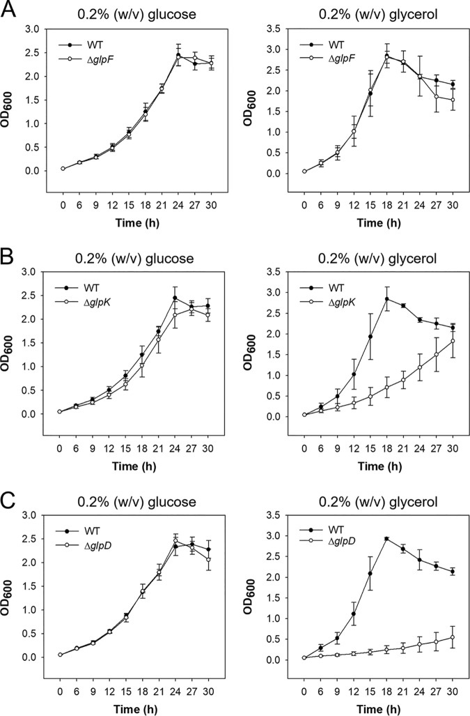
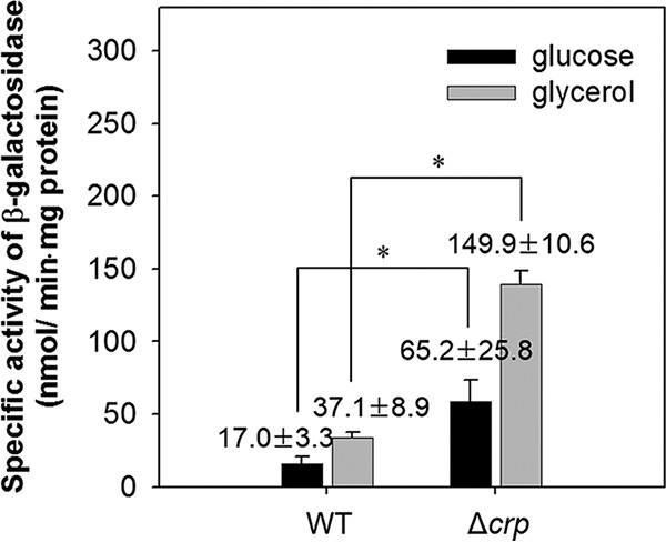
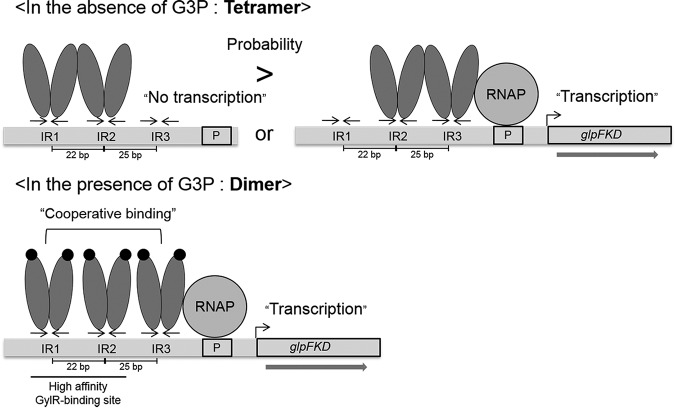
The glpD (MSMEG_6761) gene encoding glycerol-3-phosphate dehydrogenase was shown to be crucial for M. smegmatis to utilize glycerol as the sole carbon source. The glpD gene likely forms the glpFKD operon together with glpF and glpK, encoding a glycerol facilitator and glycerol kinase, respectively. The gylR (MSMEG_6757) gene, whose product belongs to the IclR family of transcriptional regulators, was identified 182 bp upstream of glpF It was demonstrated that GylR serves as a transcriptional activator and is involved in the induction of glpFKD expression in the presence of glycerol. Three GylR-binding sites with the consensus sequence (GKTCGRC-N3-GYCGAMC) were identified in the upstream region of glpF by DNase I footprinting analysis. The presence of glycerol-3-phosphate was shown to decrease the binding affinity of GylR to the glpF upstream region with changes in the quaternary structure of GylR from tetramer to dimer. Besides GylR, cAMP receptor protein (Crp) and an alternative sigma factor, SigF, are also implicated in the regulation of glpFKD expression. Crp functions as a repressor, while SigF induces expression of glpFKD under energy-limiting conditions. In conclusion, we suggest here that the glpFKD operon is under the tripartite control of GylR, SigF, and Crp, which enables M. smegmatis to integrate the availability of glycerol, cellular energy state, and cellular levels of cAMP to exquisitely control expression of the glpFKD operon involved in glycerol metabolism.IMPORTANCE Using genetic approaches, we first revealed that glycerol is catabolized through the glycolytic pathway after conversion to dihydroxyacetone phosphate in two sequential reactions catalyzed by glycerol kinase (GlpK) and flavin adenine dinucleotide (FAD)-containing glycerol-3-phosphate dehydrogenase (GlpD) in M. smegmatis Our study also revealed that in addition to the GylR transcriptional activator that mediates the induction of the glpFKD operon by glycerol, the operon is regulated by SigF and Crp, which reflect the cellular energy state and cAMP level, respectively.

Keywords: GylR transcriptional regulator; Mycobacterium; SigF; cAMP receptor protein; glycerol catabolism; glycerol kinase; glycerol-3-phosphate dehydrogenase; regulation of gene expression.


Figures

FIG 1
FIG 1
Genetic organization of the glpFKD locus in M. smegmatis mc2 155 and the upstream sequence of the glpFKD operon encompassing its promoter region and putative cis-acting elements involved in the regulation of the glpFKD operon. (A) The lengths of the overlapping and intergenic regions are given as the nucleotide numbers above the schematic diagram. (B) Four inverted repeats (IR1, IR2, IR3, and putative Crp-binding site) are marked by the head-facing arrows above their sequences. The putative promoter region of the glpFKD operon is boxed. The transcriptional start point (+1) of the glpFKD operon deduced from RNA deep-sequencing data from M. smegmatis is shaded in gray. The start codons of glpF and gylR are underlined, and the arrows above them indicate the transcriptional direction. The numbers to the left of the sequences indicate the positions of the leftmost nucleotides relative to the glpF gene. RBS, ribosome-binding site.
FIG 2
FIG 2
Growth curves of the wild-type, ΔglpF, ΔglpK, and ΔglpD mutant strains of M. smegmatis. The ΔglpF (A), ΔglpK (B), and ΔglpD (C) mutant strains, as well as the wild-type (WT) strain as the control, were grown aerobically in 7H9 medium supplemented with 0.2% (wt/vol) glucose or 0.2% (wt/vol) glycerol as a carbon source at 37°C. All values provided are the averages of the results from three biological replicates. The error bars indicate the standard deviations.
FIG 3
FIG 3
Expression of the glpFKD operon in the wild-type and ΔgylR strains of M. smegmatis. The glpF promoter activity was determined in the wild-type (WT) and ΔgylR mutant strains carrying both pMV306 and the glpF::lacZ transcriptional fusion plasmid pNCglpF. For complementation of the ΔgylR mutant, pMV306gylR (a pMV306-derived plasmid carrying the intact gylR gene and its own promoter) was introduced into the mutant (ΔgylR + gylR) in place of pMV306. The strains were grown aerobically to an OD600 of 0.45 to 0.5 in 7H9-glycerol or 7H9-glucose. Cell-free crude extracts were used to measure β-galactosidase activity. All values provided are the averages of the results from three biological replicates. The error bars indicate the standard deviations. Statistical significance was determined by two-tailed Student's t test. *, P < 0.01.
FIG 4
FIG 4
Binding of GylR to the glpFKD regulatory region. (A) DNase I footprinting analysis of the glpFKD regulatory region bound by GylR. The gylR-glpF intergenic DNA fragments containing the glpF coding strand labeled with TAMRA at their 5′ ends were incubated with increasing concentrations (0, 1.5, and 3.0 nmol) of purified GylR and then subjected to DNase I footprinting reactions. The amounts of GylR protein used are given below the lanes. The regions protected by GylR and the GylR-binding sites (IR1, IR2, and IR3) are marked by the black bar and two head-facing arrows on the right, respectively. The asterisks indicate the hypersensitive sites resulting from GylR binding. Lanes G, A, T, and C represent the sequence ladders. (B) EMSA showing the binding of purified GylR to the glpF regulatory region. The mixtures of 237-bp DNA fragments (50 fmol corresponding to 7.7 ng; specific DNA) containing the regulatory region of the glpFKD operon and 120-bp DNA fragments (50 fmol [corresponding to 3.9 ng]; control DNA) without the GylR-binding site were incubated with increasing amounts of purified GylR in the absence and presence of 50 mM G3P. The concentrations of used GylR are given above the lanes. The GylR-DNA reaction mixtures were subjected to native PAGE. After electrophoresis, the gel was stained with SYBR green EMSA gel staining solution.
FIG 5
FIG 5
Determination of the molecular mass of GylR by gel filtration chromatography. Elution profiles represent the purified GylR protein on Superose 12 (10/300). The elution profiles of purified GylR without treatment of G3P (control) and with treatment of 10 mM, 20 mM, and 50 mM G3P are indicated. β-Amylase (200 kDa), bovine serum albumin (BSA; 66 kDa), and carbonic anhydrase (CA; 29 kDa) were used as standard proteins.
FIG 6
FIG 6
Effect of deletions and mutations in the putative GylR-binding sites (IR1, IR2, and IR3) on expression of the glpFKD operon. The glpF promoter activity was determined using the pNCglpF-derived glpF::lacZ transcriptional fusions containing serial deletions of the glpF upstream region (pNCglpFΔIR1, pNCglpFΔIR12, pNCglpFΔIR123) (A) or mutations (pNCglpFM2, pNCglpFM3) in the GylR-binding sites (B). As a control (Con), pNCglpF was included in the experiment. The schematic diagrams depicting the transcriptional fusions are presented on the right. The inverted repeats of the GylR-binding sites are indicated by the head-facing arrows. Mutations within IR2 and IR3 are indicated by the asterisks. Cells of the wild-type strain of M. smegmatis harboring the transcriptional fusion plasmids were grown aerobically to an OD600 of 0.45 to 0.5 in 7H9-glycerol or 7H9-glucose. Cell-free crude extracts were used to measure β-galactosidase activity. All values provided are the averages of the results from three biological replicates. The error bars indicate the standard deviations. Statistical significance was determined by two-tailed Student's t test. *, P < 0.05.
FIG 7
FIG 7
Expression of the glpFKD operon in the wild-type and Δcrp mutant strains of M. smegmatis. The glpF promoter activity was determined using pMV306lacZglpF. The wild-type (WT) and ΔcrpMSMEG_6189) mutant strains of M. smegmatis were grown aerobically to an OD600 of 0.45 to 0.5 in 7H9-glycerol or 7H9-glucose. Cell-free crude extracts were used to measure β-galactosidase activity. All values provided are the averages of the results from three biological replicates. The error bars indicate the standard deviations. Statistical significance was determined by two-tailed Student's t test. *, P < 0.01.
FIG 8
FIG 8
EMSA showing the binding of purified Crp to the glpFKD regulatory region. Incubations of 237-bp DNA fragments containing the regulatory region of the glpFKD operon (50 fmol, corresponding to 7.7 ng; specific DNA) and 120-bp DNA fragments without the Crp-binding site (50 fmol, corresponding to 3.9 ng; control DNA) were performed with various concentrations of purified Crp (MSMEG_6189). The concentrations of Crp are given above the lanes. The Crp-DNA reaction mixtures were subjected to native PAGE, and the gel was stained with SYBR green EMSA gel staining solution.
FIG 9
FIG 9
Expression of the glpFKD operon in the ΔsigF and Δaa3 mutant strains of M. smegmatis. The glpF promoter activity was determined in the ΔsigF (A) and Δaa3 (B) mutant strains of M. smegmatis using pNCglpF. The wild-type (WT) strain with pNCglpF was included in the assay as a reference for comparison. The WT and mutant strains were grown aerobically to an OD600 of 0.45 to 0.5 in 7H9-glucose medium. Cell-free crude extracts were used to measure β-galactosidase activity. All values provided are the averages of the results from three biological replicates. The error bars indicate the standard deviations. Statistical significance was determined by two-tailed Student's t test. *, P < 0.01.
FIG 10
FIG 10
Model for the regulation of the glpFKD operon by GylR. The GylR monomers are represented by gray ovals, and the GylR-binding sites (IR1, IR2, and IR3) are marked by the head-facing arrows. The numbers between the two adjacent GylR-binding sites indicate the distances between their centers. G3P is depicted by black circles. The promoter region (P) of the glpFKD operon is boxed. RNAP, RNA polymerase.

References

    1. Blötz C, Stulke J. 2017. Glycerol metabolism and its implication in virulence in Mycoplasma. FEMS Microbiol Rev 41:640–652. doi:10.1093/femsre/fux033. - DOI - PubMed
    1. de Carvalho LP, Fischer SM, Marrero J, Nathan C, Ehrt S, Rhee KY. 2010. Metabolomics of Mycobacterium tuberculosis reveals compartmentalized co-catabolism of carbon substrates. Chem Biol 17:1122–1131. doi:10.1016/j.chembiol.2010.08.009. - DOI - PubMed
    1. Muñoz-Elías EJ, McKinney JD. 2005. Mycobacterium tuberculosis isocitrate lyases 1 and 2 are jointly required for in vivo growth and virulence. Nat Med 11:638–644. doi:10.1038/nm1252. - DOI - PMC - PubMed
    1. Youmans GP, Youmans AS. 1953. Studies on the metabolism of Mycobacterium tuberculosis. I. The effect of carbohydrates and alcohols on the growth of Mycobacterium tuberculosis var. hominis. J Bacteriol 65:92–95. - PMC - PubMed
    1. Ratledge C. 1976. The physiology of the mycobacteria. Adv Microb Physiol 13:115–244. doi:10.1016/S0065-2911(08)60039-9. - DOI - PubMed

Publication types

MeSH terms