. 2019 Oct 29;116(44):22252-22261.
doi: 10.1073/pnas.1909504116. Epub 2019 Sep 30.

Inherent reactivity of unselected TCR repertoires to peptide-MHC molecules

Affiliations

Inherent reactivity of unselected TCR repertoires to peptide-MHC molecules

S Harsha Krovi et al. Proc Natl Acad Sci U S A. .

Abstract

The repertoire of αβ T cell antigen receptors (TCRs) on mature T cells is selected in the thymus where it is rendered both self-tolerant and restricted to the recognition of major histocompatibility complex molecules presenting peptide antigens (pMHC). It remains unclear whether germline TCR sequences exhibit an inherent bias to interact with pMHC prior to selection. Here, we isolated TCR libraries from unselected thymocytes and upon reexpression of these random TCR repertoires in recipient T cell hybridomas, interrogated their reactivities to antigen-presenting cell lines. While these random TCR combinations could potentially have reacted with any surface molecule on the cell lines, the hybridomas were stimulated most frequently by pMHC ligands. The nature and CDR3 loop composition of the TCRβ chain played a dominant role in determining pMHC-reactivity. Replacing the germline regions of mouse TCRβ chains with those of other jawed vertebrates preserved reactivity to mouse pMHC. Finally, introducing the CD4 coreceptor into the hybridomas increased the proportion of cells that could respond to pMHC ligands. Thus, αβ TCRs display an intrinsic and evolutionary conserved bias for pMHC molecules in the absence of any selective pressure, which is further strengthened in the presence of coreceptors.

Keywords: T cell antigen receptor; T cell selection; major histocompatibility complex.


Conflict of interest statement

The authors declare no competing interest.

Figures

Fig. 1.
Fig. 1.
Cloned unselected TRAV libraries are diverse. Plots displaying the diversity for each of the TRAV libraries generated. In every plot, each circle represents 1 specific clone (as determined by TRAV/TRAJ usage and a unique CDR3 sequence). The size of the circle represents the frequency with which the clone is represented in the library. Finally, the color of the circle corresponds to the TRAJ gene utilized by the clone. (Top) Plots for all of the sequences in each of the 5 TRAV libraries. (Middle) Plots for only the functional sequences (sequences that are in-frame and lack stop codons) in each of the 5 TRAV libraries. (Bottom) Plots for the functional sequences recovered from transduced hybridomas for each of the 5 TRAV libraries.
Fig. 2.
Fig. 2.
Hybridomas with the NFAT-GFP reporter respond in a dose-dependent manner upon stimulation. Hybridomas expressing the B3K506 TCR (specific for the 3K peptide presented by the I-Ab MHC-II molecule) were stimulated at different concentrations of αCD3/αCD28 (A) or by the H-2b haplotype bearing CHB-2.4.4 cells with various 3K peptide concentrations (B) for 18 to 24 h. Subsequently, they were stained with an antibody targeting TCRβ and the proportion of GFP+ cells was assessed. Summary of the results from the antibody stimulation or from the antigen presentation assay with plots depicting the proportion of GFP+ cells, the geometric mean fluorescent intensity (gMFI) of the GFP+ cells, and the proportion of TCRβ+ cells as a function of stimulation dose are shown.
Fig. 3.
Fig. 3.
Unselected TRAV libraries paired to postselected TCRβ chains are MHC-reactive. (Left) TRAV libraries were paired to the 75-55β, the YAe5-62.8β, and the DO-11.10β chains and stimulated by H-2b–bearing CHB-2.4.4 cells in the presence of isotype or MHC-blocking antibodies. After 18 to 24 h of stimulation, the cells were stained with an antibody targeting TCRβ and the proportion of GFP+ cells was assessed in each stimulation condition. Results are representative of 2 independent experiments. (Right) The results are summarized for each stimulation condition with red representing the isotype antibody condition and blue representing the MHC-blocking antibody condition (***P < 0.001 by multiple t tests; error bars are mean ± SD).
Fig. 4.
Fig. 4.
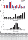
TCRβ repertoire of TCRα−/− DP thymocytes. (A) Intracellular Vβ (TRBV) staining of immature, nonsignaling DP thymocytes from C57BL/6 and TCRα−/− mice. (B) TRBJ gene usage of the sequenced TRBV13-2 rearrangements from DP thymocytes of 3 different TCRα−/− mice (error bars are mean ± SD). (C) CDR3 length distribution of the rearrangements between TRBV13-2 and TRBJ2-7 summarized for all 3 mice (error bars are mean ± SD). (D) Table depicting the 3 most frequently occurring and shared sequences. Percentages for each sequence represent the proportion of the given sequence within all TRBV13-2/TRBJ2-7 rearrangements for any given mouse.
Fig. 5.
Fig. 5.
The CD4 coreceptors strengthen intrinsic TCR specificity for MHC. (A) Bar graphs depicting the proportion of GFP+ cells after 18 to 24 h of stimulation by H-2b–bearing CHB-2.4.4 cells for TRAV libraries expressed in hybridomas bearing the labeled TCRβ chain. Red represents the isotype antibody condition and blue represents the MHC-blocking antibody condition. Each experiment was repeated 2 independent times (**P < 0.01 by multiple t tests; error bars are mean ± SD). (B) Bar graphs depicting the proportion of GFP+ cells after 18 to 24 h of stimulation by H-2b bearing CHB-2.4.4 cells for TRAV libraries expressed in hybridomas bearing the labeled TCRβ chain and expressing the CD4 coreceptor. Red represents the isotype antibody condition and blue represents the MHC-blocking antibody condition. Each experiment was repeated 2 independent times (*P < 0.05, **P < 0.01, and ***P < 0.001 by multiple t tests; error bars are mean ± SD). (C) Table displaying the location of the introduced tryptophan residue (W, in red) in the engineered TCRβ chain and how this CDR3 sequence compares with those of 2 of the frequently observed TCRβ chains. (D) Bar graphs depicting the proportion of GFP+ cells after 18 to 24 h of stimulation by H-2b–bearing CHB-2.4.4 cells for TRAV libraries expressed in hybridomas bearing the labeled TCRβ chain and either expressing the CD4 coreceptor or not. Red represents the isotype antibody condition and blue represents the MHC-blocking antibody condition. Each experiment was repeated 2 independent times (*P < 0.05, **P < 0.01, and ***P < 0.001 by multiple t tests; error bars are mean ± SD).
Fig. 6.
Fig. 6.
Cross-species conservation of reactivity between TCR and MHC molecules. (Left) The entire germline region of mouse TRBV13-2 was replaced by either frog (from the TRBV2 gene), shark (from the Vβ3-Hf73 gene), or trout (from the TRBV8S2 gene). The 13-2_3W CDR3 was coupled to each of these TCRβ chains to create chimeric TCRβ chains. These chains were then independently expressed in the hybridomas expressing the CD4 coreceptor, paired to each of the libraries and then stimulated by H-2b–bearing CHB-2.4.4 cells in the presence of isotype or MHC-blocking antibodies. After 18 to 24 h of stimulation, the cells were stained with an antibody targeting TCRβ and the proportion of GFP+ cells was assessed in each stimulation condition. Results are representative of 2 independent experiments. (Right) The results are summarized for each stimulation condition with red representing the isotype antibody condition and blue representing the MHC-blocking antibody condition (*P < 0.05, **P < 0.01, and ***P < 0.001 by multiple t tests; error bars are mean ± SD).
Fig. 7.
Fig. 7.
TCRβ libraries paired to unselected TCRα libraries react with MHC. (Left) TRBV13-2 libraries were paired to each of the libraries and expressed in hybridomas either expressing the CD4 coreceptor or not. They were then stimulated by H-2b–bearing CHB-2.4.4 cells in the presence of isotype or MHC-blocking antibodies. After 18 to 24 h of stimulation, the cells were stained with an antibody targeting TCRβ and the proportion of GFP+ cells was assessed in each stimulation condition. Results are representative of 2 independent experiments. (Right) The results are summarized for each stimulation condition with red representing the isotype antibody condition and blue representing the MHC-blocking antibody condition (*P < 0.05, **P < 0.01, and ***P < 0.001 by multiple t tests; error bars are mean ± SD).

Comment in

References

    1. Schatz D. G., Ji Y., Recombination centres and the orchestration of V(D)J recombination. Nat. Rev. Immunol. 11, 251–263 (2011). - PubMed
    1. Davis M. M., Bjorkman P. J., T-cell antigen receptor genes and T-cell recognition. Nature 334, 395–402 (1988). - PubMed
    1. Rudolph M. G., Stanfield R. L., Wilson I. A., How TCRs bind MHCs, peptides, and coreceptors. Annu. Rev. Immunol. 24, 419–466 (2006). - PubMed
    1. Marrack P., Scott-Browne J. P., Dai S., Gapin L., Kappler J. W., Evolutionarily conserved amino acids that control TCR-MHC interaction. Annu. Rev. Immunol. 26, 171–203 (2008). - PMC - PubMed
    1. La Gruta N. L., Gras S., Daley S. R., Thomas P. G., Rossjohn J., Understanding the drivers of MHC restriction of T cell receptors. Nat. Rev. Immunol. 18, 467–478 (2018). - PubMed

Publication types

Substances