. 2019 Oct 22;116(43):21641-21650.
doi: 10.1073/pnas.1910840116. Epub 2019 Sep 30.

X chromosome and autosomal recombination are differentially sensitive to disruptions in SC maintenance

Affiliations

X chromosome and autosomal recombination are differentially sensitive to disruptions in SC maintenance

Katherine Kretovich Billmyre et al. Proc Natl Acad Sci U S A. .

Abstract

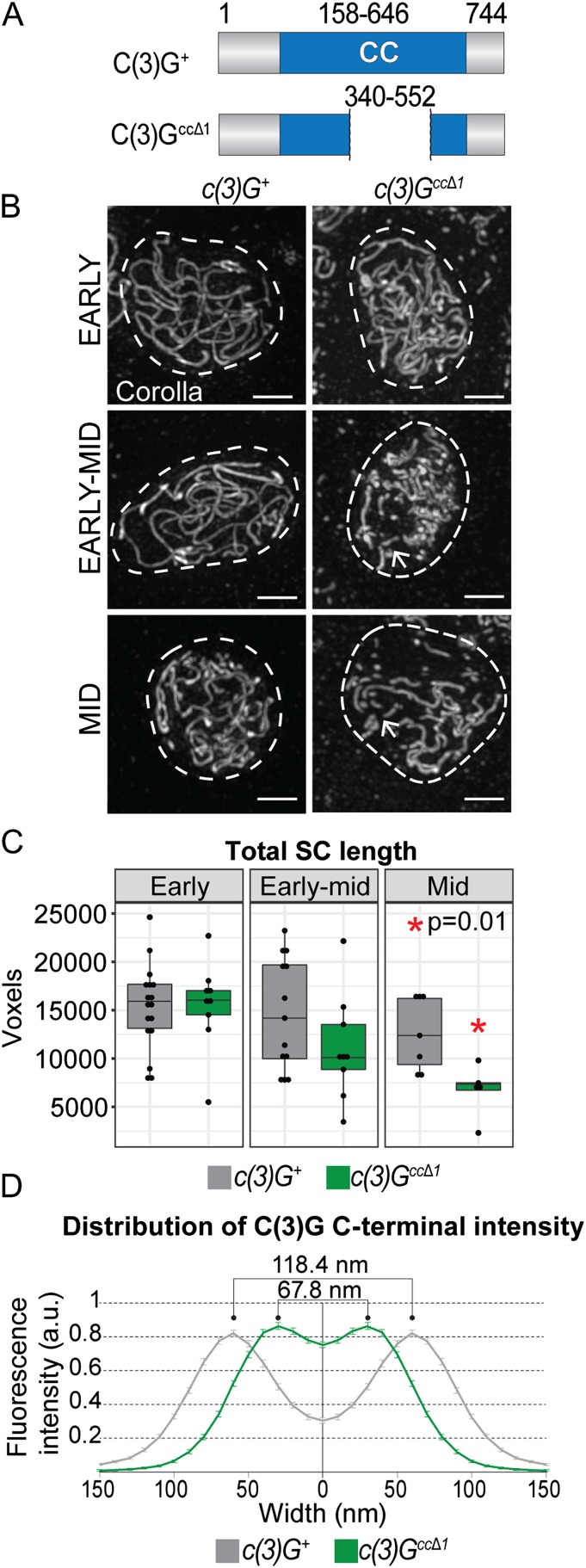
The synaptonemal complex (SC) is a conserved meiotic structure that regulates the repair of double-strand breaks (DSBs) into crossovers or gene conversions. The removal of any central-region SC component, such as the Drosophila melanogaster transverse filament protein C(3)G, causes a complete loss of SC structure and crossovers. To better understand the role of the SC in meiosis, we used CRISPR/Cas9 to construct 3 in-frame deletions within the predicted coiled-coil region of the C(3)G protein. Since these 3 deletion mutations disrupt SC maintenance at different times during pachytene and exhibit distinct defects in key meiotic processes, they allow us to define the stages of pachytene when the SC is necessary for homolog pairing and recombination during pachytene. Our studies demonstrate that the X chromosome and the autosomes display substantially different defects in pairing and recombination when SC structure is disrupted, suggesting that the X chromosome is potentially regulated differently from the autosomes.

Keywords: Drosophila; homologous recombination; meiosis; synaptonemal complex.


Conflict of interest statement

The authors declare no competing interest.

Figures

Fig. 1.
Fig. 1.
Schematic of early meiosis in Drosophila. (A) Diagram of a Drosophila germarium and SC formation (described in ref. 4). At the anterior tip of the germarium, a germline stem cell divides asymmetrically to give rise to a cystoblast, which undergoes 4 mitotic divisions with incomplete cytokinesis to yield a 16-cell cyst. At region 2A (zygotene/early pachytene), up to 4 of the 16 cells in the cyst will enter meiosis and assemble the SC (represented by blue shading) to fully synapse the chromosomes. The oocyte selection process progresses in region 2B and is characterized by 2 nuclei (pro-oocytes) with a full-length SC (early to mid pachytene) and is completed by region 3 (mid pachytene) with only 1 oocyte per cyst retaining the full-length SC and all other nuclei having backed out of the meiotic program to become nurse cells. (B) Homologous chromosome pairing and SC assembly begin at the centromeres (represented as black dots on the chromosomes) during the mitotic divisions in region 1 (28, 29). In region 2A (early pachytene) the SC (represented by blue lines) is assembled along the chromosome arms and DSBs form (orange circles). The SC is maintained along chromosome arms until stages 5 to 7 (late pachytene), when SC disassembly occurs at multiple regions along the chromosome arms. The SC persists at the centromeres into stages 8 to 9 (mid prophase) (27, 47). (C) Model of the Drosophila SC showing the transverse filament protein C(3)G (blue), central-region (CR) protein Corolla (green), central element (CE) protein CONA (black), and lateral element (LE)/cohesin proteins (gray) connected to chromatin loops (adapted from ref. 4).
Fig. 2.
Fig. 2.
In-frame deletion of part of the large coiled-coil region of C(3)G leads to a failure to maintain the SC. (A) The c(3)GccΔ1 deletion removes the amino acids 340 to 552 from the coiled-coil (CC) domain of C(3)G. The predicted protein CC is in blue [based on COILS software (38)] and gray marks the unstructured region. (B) Images showing localization of the SC protein Corolla in c(3)G+ and c(3)GccΔ1 nuclei from early pachytene (region 2A) to mid pachytene (region 3). Dashed lines indicate the location of the nucleus as defined by DAPI staining (not shown). Arrows indicate discontinuities in the SC. (Scale bars, 2 µm.) (C) Quantification of the total track length of the C(3)G–positive SC in nuclei from early, early to mid, and mid pachytene using skeleton analysis (SI Appendix). *P = 0.01 by t test. c(3)G+: n = 17 (early), n = 13 (early to mid), and n = 7 (mid); c(3)GccΔ1: n = 9 (early), n = 9 (early to mid), and n = 5 (mid). (D) The average distribution of the distance between the 2 C-terminal C(3)G tracks is shown based on a line profile analysis of STED data in each genotype (SI Appendix). The quantification resulted in an average width of 118.4 ± 0.6 nm (SEM) in wild type and 67.8 ± 0.1 nm (SEM) in c(3)GccΔ1 mutants. The average distribution was generated by averaging 46 line profiles from 8 wild-type nuclei and 35 line profiles from 12 c(3)GccΔ1 nuclei.
Fig. 3.
Fig. 3.
c(3)GccΔ1 mutants exhibit chromosome-specific defects in recombination. Recombination in c(3)GccΔ1 females on the X chromosome (A), second chromosome (B), and third chromosome (C) is plotted with the percentage of wild type on the y axis vs. chromosome location (in cM) on the x axis. Brackets along the x axis indicate truncation of that region of the chromosome. The red dashed lines mark wild-type levels of recombination and are set at 100%. P values were obtained using a Fisher’s exact test (SI Appendix, Tables S1–S3 for N values). See SI Appendix for the recessive markers used to assay recombination. For reference, below each chart is a diagram of the corresponding chromosome being analyzed displaying the relative cytological positions of the recombination markers and the approximate amounts of pericentromeric heterochromatin estimated from ref. (the black circles represent the centromere).
Fig. 4.
Fig. 4.
Two smaller in-frame deletions within the putative c(3)G coiled-coil region cause varying levels of SC defects. (A) Diagrams of the C(3)G+, C(3)GccΔ1, C(3)GccΔ2, and C(3)GccΔ3 protein with the coiled-coil region marked in blue and the unstructured regions marked in gray. (B) Images showing localization of the SC protein Corolla in c(3)G+, c(3)GccΔ2, and c(3)GccΔ3 mutants from early pachytene (region 2A) to mid pachytene (region 3). Dashed lines indicate the location of the nucleus as defined by DAPI staining (not shown). (Scale bars, 2 µm.) (C) Quantification of the total length of the C(3)G–positive SC in nuclei from early, early to mid, and mid pachytene using skeleton analysis (SI Appendix). c(3)G+ controls are the same ones used in Fig. 2. *P < 0.01 and **P < 0.001 by t test. c(3)GccΔ2: n = 11 (early), n = 11 (early to mid), and n = 7 (mid); c(3)GccΔ3: n = 10 (early), n = 10 (early to mid), and n = 5 (mid).
Fig. 5.
Fig. 5.
Loss of SC maintenance in c(3)GccΔ2 mutants in mid pachytene is not sufficient to disrupt X chromosome recombination. Recombination in c(3)GccΔ2 and c(3)GccΔ3 females on the X chromosome (A and B) and the third chromosome (C and D) is plotted with the percentage of wild type on the y axis vs. chromosome location (in cM) on the x axis. Brackets along the x axis indicate truncation of that region of the chromosome. The red dashed lines mark wild-type levels of recombination and is set at 100%. P values were obtained using a Fisher’s exact test (see SI Appendix, Tables S1 and S3 for N values). See SI Appendix for the recessive markers used to assay recombination. For reference, below each chart is a diagram of the corresponding chromosome being analyzed displaying the relative cytological positions of the recombination markers and the approximate amounts of pericentromeric heterochromatin estimated from ref. (the black circles represent the centromere).
Fig. 6.
Fig. 6.
SC in early to mid pachytene maintains homologous chromosome pairing. Fraction of paired euchromatic regions in c(3)G+ controls (gray lines), c(3)GccΔ1 (green lines), c(3)GccΔ2 (orange lines), c(3)GccΔ3 (blue lines), and c(3)Gnull flies (black lines) assessed by FISH using BAC probes against either distal or proximal euchromatin regions on the X chromosome (A) and distal, medial, or proximal euchromatin regions on the third chromosome (B) at early (E), early to mid (E-M), or mid (M) pachytene. Early pachytene is not assessed in c(3)Gnull flies due to the lack of an SC, which is the only marker to identify early pachytene nuclei. For reference, below each chart is a diagram of the corresponding chromosome being analyzed (the black circles represent the centromere). For N values, see SI Appendix, Table S5; for the distance between unpaired foci, see SI Appendix, Fig. S4 and Table S6.
Fig. 7.
Fig. 7.
Summary of SC morphology and model of the requirement for the SC in recombination and pairing maintenance. (A) Summary of SC phenotypes in c(3)G+ (gray line), c(3)GccΔ1 (green line), c(3)GccΔ2 (orange line), and c(3)GccΔ3 (blue line) flies. c(3)GccΔ1 flies displayed SC defects in early to mid pachytene while c(3)GccΔ2 flies lost the SC in mid pachytene. c(3)GccΔ3 flies never fully assembled the SC. Dotted lines indicate defects in total SC length and fragmentation. (B) A model of the requirement of full-length SC (black lines) at different stages of pachytene. Based on our data, we propose that a full-length SC is important for proper autosomal crossover (CO) placement, X chromosome recombination, and maintenance of pairing at different stages of early to mid pachytene. The gray line represents a potential role for a full-length SC that cannot be confirmed with our data.

References

    1. Hassold T., Hall H., Hunt P., The origin of human aneuploidy: Where we have been, where we are going. Hum. Mol. Genet. 16, R203–R208 (2007). - PubMed
    1. Keeney S., Giroux C. N., Kleckner N., Meiosis-specific DNA double-strand breaks are catalyzed by Spo11, a member of a widely conserved protein family. Cell 88, 375–384 (1997). - PubMed
    1. McKim K. S., Hayashi-Hagihara A., mei-W68 in Drosophila melanogaster encodes a Spo11 homolog: Evidence that the mechanism for initiating meiotic recombination is conserved. Genes Dev. 12, 2932–2942 (1998). - PMC - PubMed
    1. Hughes S. E., Miller D. E., Miller A. L., Hawley R. S., Female meiosis: Synapsis, recombination, and segregation in Drosophila melanogaster. Genetics 208, 875–908 (2018). - PMC - PubMed
    1. Nicklas R. B., Chromosome segregation mechanisms. Genetics 78, 205–213 (1974). - PMC - PubMed

Publication types

MeSH terms

Substances

LinkOut - more resources