. 2019 Sep 30;10(10):730.
doi: 10.1038/s41419-019-1965-7.

Dissociation of mitochondrial HK-II elicits mitophagy and confers cardioprotection against ischemia

Affiliations

Dissociation of mitochondrial HK-II elicits mitophagy and confers cardioprotection against ischemia

Valerie P Tan et al. Cell Death Dis. .

Abstract

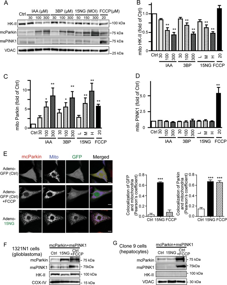
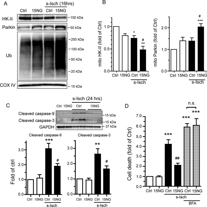
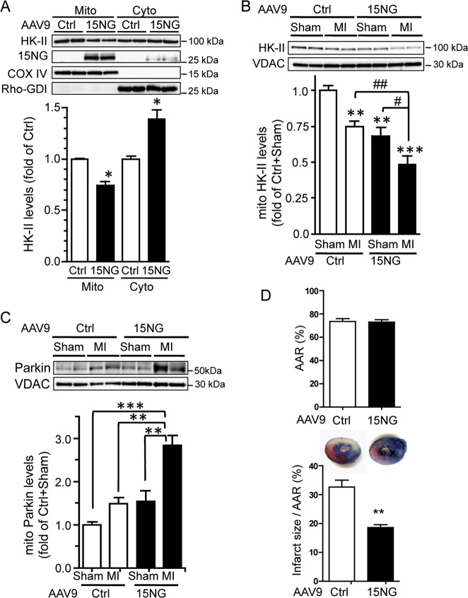
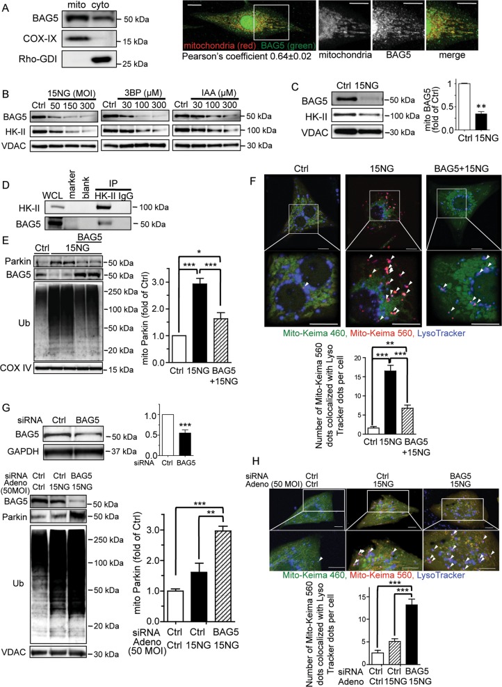
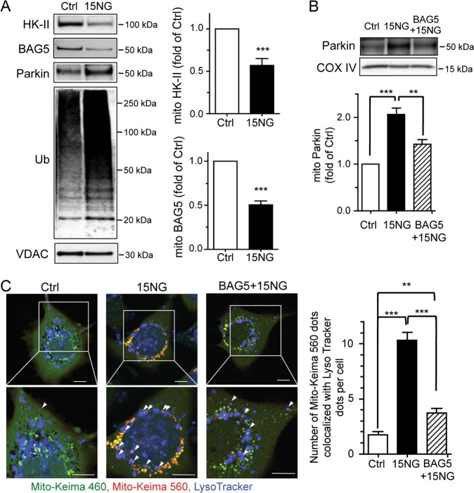
Preservation of mitochondrial integrity is critical for maintaining cellular homeostasis. Mitophagy is a mitochondria-specific type of autophagy which eliminates damaged mitochondria thereby contributing to mitochondrial quality control. Depolarization of the mitochondrial membrane potential is an established mechanism for inducing mitophagy, mediated through PINK1 stabilization and Parkin recruitment to mitochondria. Hexokinase-II (HK-II) which catalyzes the first step in glucose metabolism, also functions as a signaling molecule to regulate cell survival, and a significant fraction of cellular HK-II is associated with mitochondria (mitoHK-II). We demonstrate here that pharmacological interventions and adenoviral expression of a mitoHK-II dissociating peptide which reduce mitoHK-II levels lead to robust increases in mitochondrial Parkin and ubiquitination of mitochondrial proteins in cardiomyocytes and in a human glioblastoma cell line 1321N1, independent of mitochondrial membrane depolarization or PINK1 accumulation. MitoHK-II dissociation-induced mitophagy was demonstrated using Mito-Keima in cardiomyocytes and in 1321N1 cells. Subjecting cardiomyocytes or the in vivo heart to ischemia leads to modest dissociation of mitoHK-II. This response is potentiated by expression of the mitoHK-II dissociating peptide, which increases Parkin recruitment to mitochondria and, importantly, provides cardioprotection against ischemic stress. These results suggest that mitoHK-II dissociation is a physiologically relevant cellular event that is induced by ischemic stress, the enhancement of which protects against ischemic damage. The mechanism which underlies the effects of mitoHK-II dissociation can be attributed to the ability of Bcl2-associated athanogene 5 (BAG5), an inhibitor of Parkin, to localize to mitochondria and form a molecular complex with HK-II. Overexpression of BAG5 attenuates while knockdown of BAG5 sensitizes the effect of mitoHK-II dissociation on mitophagy. We suggest that HK-II, a glycolytic molecule, can function as a sensor for metabolic derangements at mitochondria to trigger mitophagy, and modulating the intracellular localization of HK-II could be a novel way of regulating mitophagy to prevent cell death induced by ischemic stress.


Conflict of interest statement

The authors declare that they have no conflict of interest.

Figures

Fig. 1
Fig. 1. HK-II dissociation from mitochondria induces mitochondrial localization of exogenously expressed Parkin but not PINK1.
ad mCherry-tagged Parkin (mcParkin) and miniSOG-tagged PINK1 (msPINK1) were adenovirally expressed in neonatal rat ventricular myocytes (NRVMs). Cells were treated with 3-bromopyruvate (3BP), iodoacetate (IAA), or FCCP for 1 h or infected with adenovirus encoding 15NG at 50 (L), 150 (M) or 300 (H) MOI for 18 h. Mitochondrial fractions were isolated and subjected to Western blot. bd Quantitative analysis of HK-II, mcParkin and msPINK1 levels in mitochondrial fractions. *, **p < 0.05, 0.01 versus control (Ctrl); n > 5. e. NRVMs were infected with mcParkin adenovirus and control (GFP) or 15NG adenovirus and subjected to live-cell imaging using confocal microscopy. Parkin (red); mitochondria (blue); GFP (green). Scale bars: 10 μm. Pearson’s coefficient was measured from images. n > 30, ***p < 0.001 versus ctrl. f, g mcParkin and msPINK1 were adenovirally expressed in 1321N1 cells or clone 9 hepatocytes. 15NG was expressed at 150 MOI. COX-IV or VDAC were used as mitochondrial markers as well as loading controls
Fig. 2
Fig. 2. HK-II dissociation induced by 15NG expression leads to mitochondrial localization of Parkin but not PINK1 in NRVMs.
a PINK1 Western blot was carried out on mitochondrial fractions isolated from NRVMs treated with FCCP (20 μM for 14 h) or 15NG (150 MOI for 18 h). Middle panels show TMRE fluorescence images using confocal microscopy and quantification of cellular TMRE fluorescence intensity. Scale bars: 10 μm. n > 60 from three independent experiments. Right panel shows cellular ATP levels in Ctrl and 15NG expressing cells. n = 5. b Mitochondrial and cytosolic fractions were subjected to Western blot for Parkin and ubiquitin (Ub). COX-IV and Rho-GDI were used as mitochondrial and cytosolic markers respectively. **p < 0.01 versus control (Ctrl); n = 4. c, d. Cells were transfected with siRNA against PINK1 or Parkin. PINK1 and Parkin levels were assessed by Western blot in whole cell lysates. α-actinin was used as a loading control. e, f NRVMs transfected with siRNA against PINK1 or Parkin were infected with control or 15NG adenovirus and mitochondrial fractions were subjected to Western blot. g, h. NRVMs were infected with adenovirus encoding control (GFP) or 15NG, with or without adenovirus encoding wild-type HK-II (WT HK-II, 100 MOI) or the mitochondria binding-deficient mutant HK-II (ΔN HK-II, 100 MOI). Mitochondrial fractions were subjected to Western blot. **, ***p < 0.01, p < 0.001; n = 5. i Parkin was visualized by mCherry fluorescence (red in the merged images) and mitochondria were visualized by MitoTracker Deep Red (blue in the merged images). Scale bars: 10 μm. Pearson’s coefficient was measured from images, n > 30 from four independent experiments., ***p < 0.001 versus Ctrl
Fig. 3
Fig. 3. MitoHK-II dissociation induces mitophagy.
a LC3 Western blot was carried out in mitochondrial and cytosolic fractions isolated from NRVMs expressing control (Ctrl) or 15NG (150 MOI for 18 h). **p < 0.01 versus Ctrl; n = 8. b To assess mitophagy, Mito-Keima was adenovirally co-expressed in NRVMs with Ctrl or 15NG for 32 h. LysoTracker Blue (250 nM) was loaded onto cells for 1 h prior to visualization. FCCP was used as a positive control (20 μM for 16–20 h). Scale bars: 10 μm. Arrow heads indicate the purple dots showing both MitoKeima fluorescence excited at 560 nm and LysoTracker Blue fluorescence. Data are quantified in (c); n > 40, from four independent experiments. ***p < 0.001 versus Ctrl. d After 48 h infection, cells were collected and subjected to Western blotting analysis of VDAC and COX IV (mitochondrial proteins), Lamin A/C (nuclear protein) and GAPDH (loading control). The lysosome inhibitor Bafilomycin A1 (BFA) was used at 50 nM. **p < 0.01; n = 5
Fig. 4
Fig. 4. 15NG expression at low MOI (50 MOI) enhances mitoHK-II dissociation and Parkin recruitment to mitochondria during ischemia and confers protection in NRVMs.
a NRVMs were infected with control (Ctrl) or 15NG adenovirus at 50 MOI and subjected to simulated ischemia (s-Isch) for 16 h. Mitochondrial fractions were subjected to Western blot analysis for HK-II, Parkin and Ub. b Quantification of HK-II and Parkin levels in the mitochondrial fraction. *, **, ***p < 0.05, p < 0.01, p < 0.001 versus Ctrl. #p < 0.05 versus s-Isch/Ctrl; n = 5. c Western blot analysis of whole cell lysates from NRVMs for cleaved caspase-9 and cleaved caspase-3 after 24 h of s-Isch. **, ***p < 0.01, p < 0.001 versus Ctrl. #p < 0.05 versus s-Isch/Ctrl; n = 4–5. d S-Isch-induced apoptosis was assessed. ***p < 0.001 versus Ctrl. ##p < 0.01 versus s-Isch/Ctrl; n = 4–6
Fig. 5
Fig. 5. AAV9-mediated 15NG expression enhances mitoHK-II dissociation and Parkin localization to mitochondria induced by ischemia in the in vivo heart, and has a cardioprotective effect.
a AAV9-Ctrl or AAV9-15NG was injected into mouse via the tail vein, and 2 wks later, mitochondrial and cytosolic fractions were isolated from the heart for Western blot analysis. *, **p < 0.05, p < 0.01 versus Ctrl; n = 4–5. b, c Mice were injected with AAV9-Ctrl or AAV9-15NG for 2 wks, subjected to sham surgery or regional ischemia by ligation of the left anterior descending (LAD) artery (LAD ligation; MI surgery) for 1 h, and mitochondrial fractions were isolated from the heart. b Western blot for HK-II in the mitochondrial fraction. **, ***p < 0.01, p < 0.001 versus Ctrl + Sham. #, ##, p < 0.05, p < 0.01; n = 8–12. c Western blot for Parkin in the mitochondrial fraction. **, ***p < 0.01, p < 0.001; n = 8–12. d Mice injected with AAV9-Ctrl or AAV9-15NG for 2 wks were subjected to LAD ligation for 2 h, Area at Risk (AAR) and infarct size were determined by Evans blue and TTC respectively, and percentage of infarct size per AAR was calculated. **p < 0.01; n = 10–12
Fig. 6
Fig. 6. BAG5 forms an immunocomplex with HK-II and is dissociated from mitochondria by 15NG expression, contributing to 15NG-induced mitophagy in NRVMs.
a Mitochondrial and cytosolic fractions were isolated from NRVMs for Western blot for BAG5. Immunofluorescence images show BAG5 colocalizing with mitochondria (MitoTracker Red). b NRVMs were subjected to various interventions to dissociate mitoHK-II (see Fig. 1 legend), and BAG5 and HK-II levels in the mitochondrial fraction were determined by Western blot. c Quantification of BAG5 levels in NRVMs expressing GFP (Ctrl) or 15NG. **p < 0.01 versus Ctrl; n = 10. d HK-II was immunoprecipitated from NRVMs and subjected to Western blot for BAG5. e BAG5 was adenovirally co-expressed in NRVMs with GFP (Ctrl) or 15NG and mitochondrial fractions were isolated and subjected to Western blot for Parkin, BAG5, Ub and VDAC. *, ***p < 0.05, p < 0.001; n = 5–7. f Mito-Keima assay in NRVMs expressing GFP (Ctrl) or 15NG with or without BAG5 co-expression. Arrow heads indicate double-positive fluorescence by both MitoKeima excited at 560 nm and LysoTracker Blue, seen as purple dots. Scale bars: 10 μm. The number of purple dots per cell were quantified. **, ***p < 0.01, p < 0.001; n = 50–70 from four independent experiments. g BAG5 was knocked-down by siRNA in NRVMs. ***p < 0.001 versus ctrl, n = 3. After 48 h transfection, cells were treated with Ctrl or 15NG adenovirus at low MOI (50 MOI), and mitochondrial fractions were isolated and subjected to Western blot for BAG5, Parkin, Ub and VDAC. **, ***p < 0.01, p < 0.001; n = 3-4. h Mito-Keima assay in NRVMs subjected to BAG5 knockdown and 15NG expression (50 MOI). Scale bars: 10 μm. ***p < 0.001, n > 45 from three independent experiments
Fig. 7
Fig. 7. HK-II dissociation decreases mitochondrial BAG5 association and induces Parkin-mediated mitophagy in a human glioblastoma cell line 1321N1.
a 1321N1 cells were infected with GFP (Ctrl) or 15NG adenovirus for 24 h, and mitochondrial fractions were isolated and subjected to Western blot for HK-II, BAG5, Parkin, Ub, and VDAC. ***p < 0.001 versus ctrl, n = 4. b BAG5 was adenovirally co-expressed in 1321N1 cells with GFP (Ctrl) or 15NG, and mitochondrial fractions were isolated and subjected to Western blot for Parkin and COX IV. **, ***p < 0.01, p < 0.001; n = 4–8. c Mito-Keima assay in 1321N1 cells expressing Ctrl or 15NG with or without BAG5 co-expression. Arrow heads indicate double-positive fluorescence by both MitoKeima excited at 560 nm and LysoTracker Blue, seen as purple dots. Scale bars: 10 μm. The number of purple dots per cell were quantified. **, ***p < 0.01, p < 0.001; n = 65–85 from four independent experiments
Fig. 8
Fig. 8
HK-II dissociation from mitochondria triggers Parkin-mediated mitophagy in response to metabolic suppression

References

    1. Whelan RS, Kaplinskiy V, Kitsis RN. Cell death in the pathogenesis of heart disease: mechanisms and significance. Annu. Rev. Physiol. 2010;72:19–44. doi: 10.1146/annurev.physiol.010908.163111. - DOI - PubMed
    1. Dillon CP, Green DR. Molecular cell biology of apoptosis and necroptosis in cancer. Adv. Exp. Med. Biol. 2016;930:1–23. doi: 10.1007/978-3-319-39406-0_1. - DOI - PubMed
    1. Bravo-San Pedro JM, Kroemer G, Galluzzi L. Autophagy and mitophagy in cardiovascular disease. Circ. Res. 2017;120:1812–1824. doi: 10.1161/CIRCRESAHA.117.311082. - DOI - PubMed
    1. Nah J, Miyamoto S, Sadoshima J. Mitophagy as a protective mechanism against myocardial stress. Compr. Physiol. 2017;7:1407–1424. doi: 10.1002/cphy.c170005. - DOI - PubMed
    1. Youle RJ, Narendra DP. Mechanisms of mitophagy. Nat. Rev. Mol. Cell Biol. 2011;12:9–14. doi: 10.1038/nrm3028. - DOI - PMC - PubMed

Publication types