Review
. 2020 Jan;20(1):25-39.
doi: 10.1038/s41577-019-0218-4. Epub 2019 Sep 30.

Tumour-intrinsic resistance to immune checkpoint blockade

Affiliations
Review

Tumour-intrinsic resistance to immune checkpoint blockade

Anusha Kalbasi et al. Nat Rev Immunol. 2020 Jan.

Abstract

'Immune checkpoint blockade' for cancer describes the use of therapeutic antibodies that disrupt negative immune regulatory checkpoints and unleash pre-existing antitumour immune responses. Antibodies targeting the checkpoint molecules cytotoxic T lymphocyte antigen 4 (CTLA4), programmed cell death 1 (PD1) and PD1 ligand 1 (PD-L1) have had early success in the clinic, which has led to approval by the US Food and Drug Administration of multiple agents in several cancer types. Yet, clinicians still have very limited tools to discriminate a priori patients who will and will not respond to treatment. This has fuelled a wave of research into the molecular mechanisms of tumour-intrinsic resistance to immune checkpoint blockade, leading to the rediscovery of biological processes critical to antitumour immunity, namely interferon signalling and antigen presentation. Other efforts have shed light on the immunological implications of canonical cancer signalling pathways, such as WNT-β-catenin signalling, cell cycle regulatory signalling, mitogen-activated protein kinase signalling and pathways activated by loss of the tumour suppressor phosphoinositide phosphatase PTEN. Here we review each of these molecular mechanisms of resistance and explore ongoing approaches to overcome resistance to immune checkpoint blockade and expand the spectrum of patients who can benefit from immune checkpoint blockade.


Conflict of interest statement

Competing interests

A.K. has no competing financial or other conflicts of interest. A.R. has received honoraria from consulting with Amgen, Bristol-Myers Squibb, Chugai, Genentech, Merck, Novartis and Roche and is or has been a member of the scientific advisory board and holds stock in Advaxis, Arcus Biosciences, Bioncotech Therapeutics, Compugen, CytomX, Five Prime, FLX-Bio, ImaginAb, Isoplexis, Kite-Gilead, Lutris Pharma, Merus, PACT Pharma, Rgenix and Tango Therapeutics.

Figures

Fig. 1 |
Fig. 1 |. Interferon signalling in adaptive programmed cell death 1 ligand 1 expression.
a | A pre-existing antitumour immune response is essential for effective immune checkpoint blockade. Tumour-reactive T cells, which recognize tumour neoantigens in the context of MHC class I or class II, release interferon-γ (IFNγ), resulting in activation of the Janus kinase (JAK)– signal transducer and activator of transcription (STAT) signalling pathway. This activates the transcription factor interferon regulatory factor 1 (IRF1), which then activates the transcription of PDL1. This results in adaptive expression of programmed cell death 1 ligand 1 (PD-L1) on the surface of tumour cells, which negatively regulates the antitumour T cell response. Antibodies against PD1 or PD-L1 disrupt this negative feedback loop to restore antitumour immunity. b | A similar scenario in which tumour-specific T cells encounter antigen in the context of MHC, resulting in the release of IFNγ. However, here the IFNγ signal is not transmitted by the tumour cell owing to genetic deficiencies in the IFNγ signalling pathway (affecting, for example, JAK1 or JAK2) and adaptive PD-L1 expression does not occur. In the absence of adaptive PD-L1 expression, PD1–PD-L1 immune checkpoint blockade is ineffective. IFNγR, IFNγ receptor; TCR, T cell receptor.
Fig. 2 |
Fig. 2 |. Timeline of original discoveries of the importance of the interferon-γ (IFnγ) pathway and antigen presentation in antitumour immunity.
This timeline also highlights the rediscovery of these key pathways in the era of immune checkpoint blockade after 2011. Discoveries relating to the IFNγ pathway are shown in blue, discoveries relating to antigen presentation are shown in pink and discoveries relating to both the IFNγ pathway and antigen presentation are shown in light pink. B2M, β2-microglobulin; JAK, Janus kinase; PD1, programmed cell death 1.
Fig. 3 |
Fig. 3 |. Resistance to immune checkpoint blockade: tumour-intrinsic escape mechanisms.
a | Multiple unbiased CRISPR-based screens have uncovered the critical role of tumour-intrinsic interferon signalling in response to immune checkpoint blockade and T cell-based immunotherapy. These studies have identified components of the interferon-γ (IFNγ) and type I interferon signalling pathway such as Janus kinase 1 (JAK1), JAK2, signal transducer and activator of transcription 1 (STAT1) and IFNγ receptor I (IFNGR1) and IFNGR2 as critical to the success or failure of immune checkpoint blockade. The studies also identifed a role in responses to immune checkpoint blockade for lesser known regulators include the surface receptor apelin receptor (APLNR), which modulates upstream sensitivity to IFNγ and type I interferon signalling, tyrosine-protein phosphatase non-receptor type 2 (PTPN2), which modulates upstream sensitivity to IFNγ signalling, BRD7 and the DNA-binding subunits ARID2 and PBRM1, which are part of the chromatin remodelling complex PBAF and are involved in the regulation of IFNγ target genes; and the enzyme double-stranded RNA (dsRNA)-specific adenosine deaminase (ADAR1), which negatively regulates endogenous dsRNA levels. Type I interferon and IFNγ signalling converge on IFNγ activation sites (GAS) in the DNA and activate transcriptional regulators such as interferon regulatory factor 1 (IRF1), which then drive key outputs from interferon signalling. b | A number of studies showed that interferon-independent defects in antigen presentation can also lead to immune evasion and resistance to immune checkpoint blockade. These defects can occur in the HLA loci or in the MHC class I complex component β2-microglobulin (B2M),. Other defects can occur in the antigen processing machinery, such as the membrane-bound transporter proteins TAP1 and TAP2 and the immunoproteasome subunits PSMB8, PSMB9 or PSMB10, or in the transcriptional regulation of MHC class I (such as the cytoplasmic protein NLRC5). MHC class I expression can also be affected at the post-transcriptional level. The RNA-binding protein MEX3B can bind HLA-A transcripts, resulting in their degradation and reduced expression of MHC class I molecules. MEX3B was found to be upregulated in patients with melanoma who did not respond to checkpoint blockade. Although higher MHC class II antigen presentation on tumour cells has been associated with improved responses to immune checkpoint blockade, genetic defects in MHC class II genes or the MHC class II transcriptional activator CIITA have not been identified in cases of resistance to immune checkpoint blockade. IFNAR1, interferon-α and β receptor subunit 1; IFNR, interferon receptor; UTR, untranslated region.
Fig. 4 |
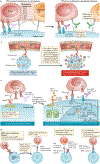
Fig. 4 |. Overcoming tumour-intrinsic resistance to immune checkpoint blockade.
a | The programmed cell death 1 (PD1)–PD1 ligand 1 (PD-L1) axis may not be the sole negative regulator of antitumour T cell responses. Alternative immune checkpoint molecules expressed on tumour cells, or myeloid cells in the tumour microenvironment, prevent effective antitumour immunity; combined immune checkpoint blockade may disrupt this resistance mechanism (right panel). b | Immunologically cold tumour types lack pre-existing antitumour T cell responses, rendering immune checkpoint blockade ineffective. Approaches to prime the immune system against tumours by causing immunogenic cell death (oncolytic viruses or cytotoxic therapy), priming antigen-presenting cells (APCs; using Toll-like receptor (TLR) agonists and CD40 agonists) or increasing tumour cell sensitivity to double-stranded RNA (dsRNA; such as inhibition of dsRNA-specific adenosine deaminase (ADAR1)) can reprogramme the immunologically cold state into a checkpoint blockade responsive state. c | Tumours without interferon-γ (IFNγ) signalling lack the capacity to adaptively express PD-L1 in response to IFNγ. In some tumours in which MHC class I antigen presentation is largely dependent on IFNγ signalling, loss of IFNγ signalling equates to loss of antigen presentation. Activation of the alternative interferon pathway (type I interferon) through TLR agonists, oncolytic viruses or other means, can also result in activation of signal transducer and activator of transcription 1 (STAT1) and STAT2 signalling, which drives transcription of PD-L1 and MHC class I via the induction of interferon regulatory factor 1 (IRF1). d | Other tumours are resistant to immune checkpoint blockade after loss of MHC class I expression via genetic alterations (such as loss of β2-microglobulin (B2M) and loss of HLA heterozygosity). Three approaches can be successful in this setting: (1) chimeric antigen receptor (CAR) T cells recognize their targets independently of MHC class I expression; (2) adoptive transfer of natural killer (NK) cells or NK cell stimulation with cytokines such as IL-2 or IL-15, as these target cells lack MHC class I expression; and (3) vaccination or adoptive T cell therapy to generate responses against a specific MHC class II-restricted antigen. B2M, β2-microglobulin; DC, dendritic cell; GAS, IFNγ activation sites; IFNAR1, interferon-α and β receptor subunit 1; IFNγR, IFNγ receptor; JAK1, Janus kinase 1; TCR, T cell receptor.
Fig. 5 |
Fig. 5 |. Oncogenic signalling pathways affecting antitumour immunity and resistance to immune checkpoint blockade.
a | Oncogenic signalling pathways provide unique tumour-intrinsic mechanisms of immune evasion. Here we highlight four key oncogenic signalling pathways implicated in antitumour immunity: the mitogen-activated protein kinase (MAPK) signalling pathway, the WNT–β-catenin pathway, the cyclin-dependent kinase 4 (CDK4)–CDK6 cell cycle signalling pathway and pathways activated as a result of loss of the phosphoinositide phosphatase PTEN. b | Therapeutic disruption of CDK4–CDK6 signalling (for example with palbociclib or abemaciclib), MAKP signalling (BRAF inhibitors) or WNT signalling can reverse the tumour-intrinsic T cell-excluded state and restore sensitivity to immune checkpoint blockade. BATF3, basic leucine zipper transcriptional factor ATF-like 3; CCL4, CC-chemokine ligand 4; DNMT1, DNA (cytosine-5)-methyltransferase 1; dsRNA, double-stranded RNA; IFNγ, interferon-γ; PAMPs, pathogen-associated molecular patterns; TNF, tumour necrosis factor; Treg cell, regulatory T cell.

References

    1. Chowell D et al. Patient HLA class I genotype influences cancer response to checkpoint blockade immunotherapy. Science 359, 582–587 (2018). - PMC - PubMed
    1. Pitt JM et al. Resistance mechanisms to immune-checkpoint blockade in cancer: tumor-intrinsic and -extrinsic factors. Immunity 44, 1255–1269 (2016). - PubMed
    1. Fessler J, Matson V & Gajewski TF Exploring the emerging role of the microbiome in cancer immunotherapy. J. Immunother. Cancer 7, 108 (2019). - PMC - PubMed
    1. Yarchoan M, Hopkins A & Jaffee EM Tumor mutational burden and response rate to PD-1 inhibition. N. Engl. J. Med 377, 2500–2501 (2017). - PMC - PubMed
    1. Rizvi NA et al. Mutational landscape determines sensitivity to PD-1 blockade in non-small cell lung cancer. Science 348, 124–128 (2015). - PMC - PubMed
    2. Association of mutational burden and sensitivity to immune checkpoint blockade has been observed across malignancies, including lung cancer. This study provides further support for the importance of tumour-intrinsic biology for sensitivity to immune checkpoint blockade.

Publication types