. 2020 Aug;19(4):1319-1329.
doi: 10.1007/s10237-019-01229-y. Epub 2019 Sep 30.

A population-specific material model for sagittal craniosynostosis to predict surgical shape outcomes

Affiliations

A population-specific material model for sagittal craniosynostosis to predict surgical shape outcomes

Alessandro Borghi et al. Biomech Model Mechanobiol. 2020 Aug.

Abstract

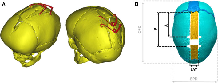
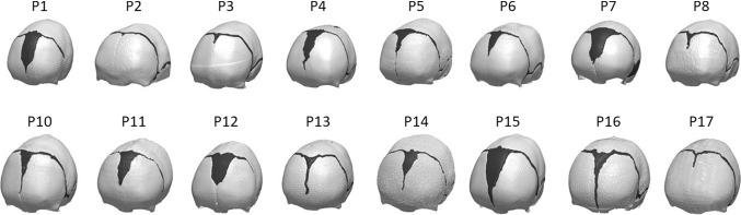
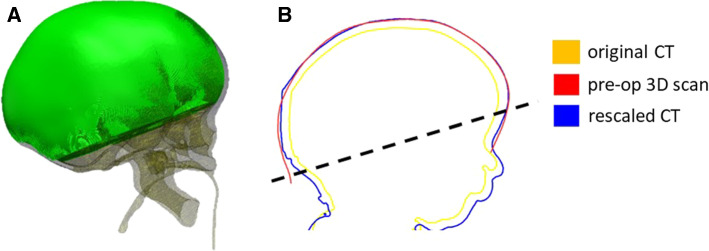
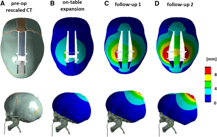
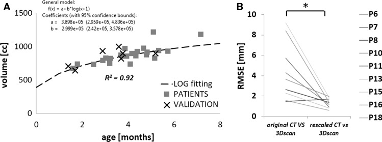
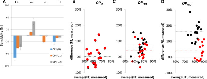
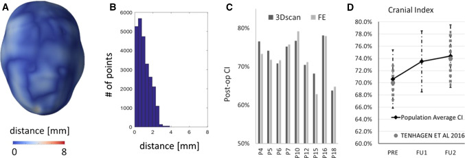
Sagittal craniosynostosis consists of premature fusion (ossification) of the sagittal suture during infancy, resulting in head deformity and brain growth restriction. Spring-assisted cranioplasty (SAC) entails skull incisions to free the fused suture and insertion of two springs (metallic distractors) to promote cranial reshaping. Although safe and effective, SAC outcomes remain uncertain. We aimed hereby to obtain and validate a skull material model for SAC outcome prediction. Computed tomography data relative to 18 patients were processed to simulate surgical cuts and spring location. A rescaling model for age matching was created using retrospective data and validated. Design of experiments was used to assess the effect of different material property parameters on the model output. Subsequent material optimization-using retrospective clinical spring measurements-was performed for nine patients. A population-derived material model was obtained and applied to the whole population. Results showed that bone Young's modulus and relaxation modulus had the largest effect on the model predictions: the use of the population-derived material model had a negligible effect on improving the prediction of on-table opening while significantly improved the prediction of spring kinematics at follow-up. The model was validated using on-table 3D scans for nine patients: the predicted head shape approximated within 2 mm the 3D scan model in 80% of the surface points, in 8 out of 9 patients. The accuracy and reliability of the developed computational model of SAC were increased using population data: this tool is now ready for prospective clinical application.

Keywords: Craniofacial surgery; Design of experiments; Finite element modelling; Scaphocephaly; Spring cranioplasty.


Figures

Fig. 1
Fig. 1
a 3D reconstruction of post-operative CT images from a SAC patient; b Surgical osteotomies and measurements recorded during surgery: A = distance between the coronal suture and the anterior spring; P = distance between the coronal suture and the posterior spring; LAT = dimension of the parasagittal osteotomy; OFD = occipitofrontal diameter; BPD = biparietal diameter
Fig. 2
Fig. 2
Patient population for the study—each model is made of skull (white) and cranial sutures (black)
Fig. 3
Fig. 3
a Visualization of the control volume VCT used for the model rescaling. b Sagittal cross section of a patient head to show comparison showing comparison between head shape retrieved from the original CT (yellow), the rescaled dataset (blue) and a pre-op 3D scan (red); the region used for the RMSE is above the dotted line
Fig. 4
Fig. 4
a Pre-operative CAD model with spring conditions, b simulated spring expansion on-table, c at follow-up 1, and d 2 for a representative patient—top view (top) and lateral view (bottom)
Fig. 5
Fig. 5
a Calvarial growth curve for sagittal craniosynostosis population (squares) with validation points (cross). b RMSE between the three head shape models showing accuracy of rescaling (* indicates statistical difference)
Fig. 6
Fig. 6
a Model sensitivity histogram from the DoE performed on nine patients. Bland–Altman plot showing a comparison between the results of simulations vs measurements in case of literature values of material properties (black) and population optimized material model (red) for b insertion (OPIO), c follow-up 1 (OPFU1), d follow-up 2 (OPFU2); results are shown in terms of percentage of nominal spring size (60 mm)
Fig. 7
Fig. 7
a Sample patient distance map between the predicted post-operative head shape for a representative patient. b Histogram showing the error distribution for the representative patient. c Bar chart showing a comparison between the post-op CI measured from 3D scan and that predicted from the FE simulations. d Population average CI measured from preoperative (PRE) models and simulated at the time of FU1 and FU2; in grey, the values of a similar population reported by Tenhagen et al. (2016)

References

    1. ANSYS 17.2. User Manual
    1. Anderson PJ, Netherway DJ, McGlaughlin K, David DJ. Intracranial volume measurement of sagittal craniosynostosis. J Clin Neurosci. 2007;14:455–458. doi: 10.1016/j.jocn.2006.07.001. - DOI - PubMed
    1. Arnaud E, Marchac A, Jeblaoui Y, et al. Spring-assisted posterior skull expansion without osteotomies. Child’s Nerv Syst. 2012;28:1545–1549. doi: 10.1007/s00381-012-1843-4. - DOI - PubMed
    1. Aung SC, Ngim RC, Lee ST. Evaluation of the laser scanner as a surface measuring tool and its accuracy compared with direct facial anthropometric measurements. Br J Plast Surg. 1995;48:551–558. doi: 10.1016/0007-1226(95)90043-8. - DOI - PubMed
    1. Beaumont CAA, Knoops PGM, Borghi A, et al. Three-dimensional surface scanners compared with standard anthropometric measurements for head shape. J Cranio-Maxillofacial Surg. 2017 doi: 10.1016/j.jcms.2017.03.003. - DOI - PubMed

LinkOut - more resources