. 2020 Jun;71(6):2050-2066.
doi: 10.1002/hep.30975. Epub 2020 Mar 16.

Probiotic Lactobacillus rhamnosus GG Prevents Liver Fibrosis Through Inhibiting Hepatic Bile Acid Synthesis and Enhancing Bile Acid Excretion in Mice

Affiliations

Probiotic Lactobacillus rhamnosus GG Prevents Liver Fibrosis Through Inhibiting Hepatic Bile Acid Synthesis and Enhancing Bile Acid Excretion in Mice

Yunhuan Liu et al. Hepatology. 2020 Jun.

Abstract

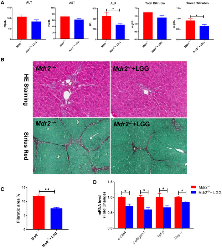
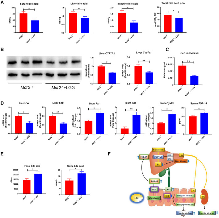
Background and aims: Cholestatic liver disease is characterized by gut dysbiosis and excessive toxic hepatic bile acids (BAs). Modification of gut microbiota and repression of BA synthesis are potential strategies for the treatment of cholestatic liver disease. The purpose of this study was to examine the effects and to understand the mechanisms of the probiotic Lactobacillus rhamnosus GG (LGG) on hepatic BA synthesis, liver injury, and fibrosis in bile duct ligation (BDL) and multidrug resistance protein 2 knockout (Mdr2-/- ) mice.

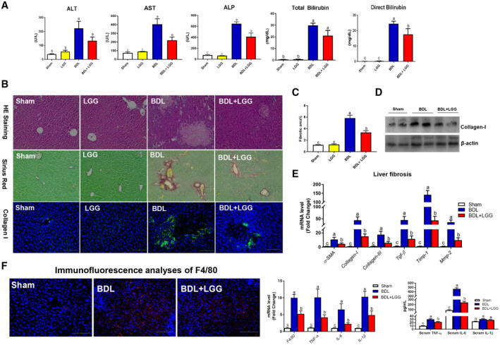
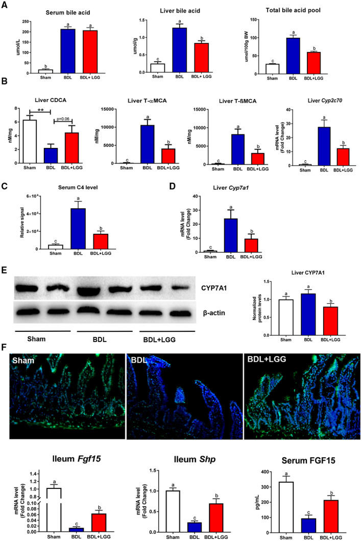
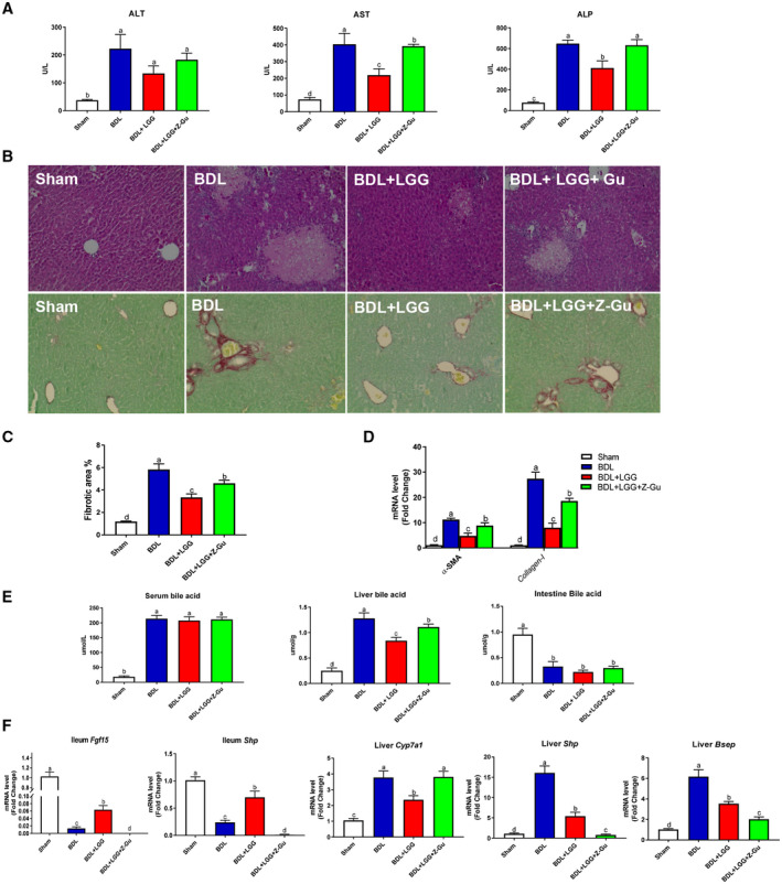
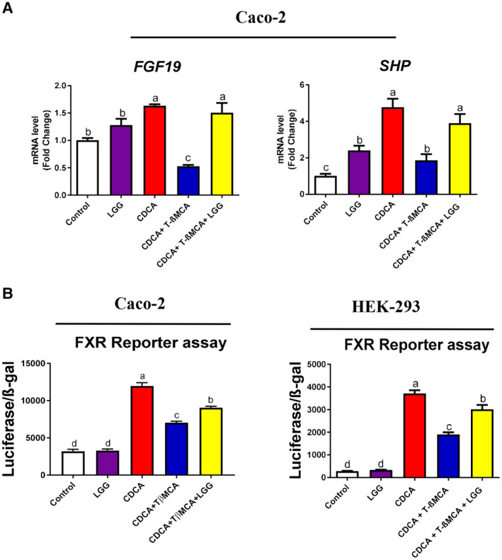
Approach and results: Global and intestine-specific farnesoid X receptor (FXR) inhibitors were used to dissect the role of FXR. LGG treatment significantly attenuated liver inflammation, injury, and fibrosis with a significant reduction of hepatic BAs in BDL mice. Hepatic concentration of taurine-β-muricholic acid (T-βMCA), an FXR antagonist, was markedly increased in BDL mice and reduced in LGG-treated mice, while chenodeoxycholic acid, an FXR agonist, was decreased in BDL mice and normalized in LGG-treated mice. LGG treatment significantly increased the expression of serum and ileum fibroblast growth factor 15 (FGF-15) and subsequently reduced hepatic cholesterol 7α-hydroxylase and BA synthesis in BDL and Mdr2-/- mice. At the molecular level, these changes were reversed by global and intestine-specific FXR inhibitors in BDL mice. In addition, LGG treatment altered gut microbiota, which was associated with increased BA deconjugation and increased fecal and urine BA excretion in both BDL and Mdr2-/- mice. In vitro studies showed that LGG suppressed the inhibitory effect of T-βMCA on FXR and FGF-19 expression in Caco-2 cells.

Conclusion: LGG supplementation decreases hepatic BA by increasing intestinal FXR-FGF-15 signaling pathway-mediated suppression of BA de novo synthesis and enhances BA excretion, which prevents excessive BA-induced liver injury and fibrosis in mice.


Figures

Figure 1
Figure 1
LGG treatment improved liver injury and fibrosis in BDL mice. (A) Serum ALT, AST, and ALP levels. Serum total bilirubin and direct bilirubin levels. (B) Representative images of liver specimens stained with hematoxylin and eosin (original magnification ×200), sirius red (×200), and immunofluorescence analyses of collagen I (×200). (C) Morphometric quantification of sirius red staining–positive area. (D) Hepatic protein expression of collagen I. (E) Hepatic mRNA expression of liver fibrosis–related genes. (F) Immunofluorescence analyses of F4/80 (×200); hepatic mRNA expression of liver inflammation‐related genes; and serum levels of TNF‐α, IL‐6, and IL‐1β. Data are expressed as mean ± SEM (n = 5‐8). Columns with different letters differ significantly (P < 0.05). Abbreviations: HE, hematoxylin and eosin; Mmp‐2, matrix metallopeptidase 2.
Figure 2
Figure 2
LGG treatment changed hepatic BA metabolism and activated intestinal FXR signaling. (A) Serum BA levels, liver total BA levels, and total BA pool. (B) Hepatic levels of CDCA, T‐αMCA, T‐βMCA, and mRNA expression of Cyp2c70. (C) Serum C4 level. (D) Hepatic mRNA expression of Cyp7a1. (E) Hepatic protein expression and quantification of CYP7A1. (F) Immunofluorescence analyses of ileum FGF‐15 (×200), ileum mRNA expression of Fgf‐15 and Shp, and serum FGF‐15 levels. Data are expressed as mean ± SEM (n = 5‐8). Columns with different letters differ significantly (P < 0.05).
Figure 3
Figure 3
Global inhibition of FXR activation abolished the protective effects of LGG. (A) Serum ALT, AST, and ALP activities. (B) Representative images of liver specimens stained with hematoxylin and eosin (×200) and sirius red (×200). (C) Morphometric quantification of sirius red staining–positive area. (D) Hepatic mRNA expression of α‐SMA and collagen I. (E) Serum BA levels, Hepatic total BA levels, and intestinal BA levels. (F) Ileum mRNA expression of Fgf‐15 and Shp and hepatic mRNA expression of Cyp7a1, Shp, and Bsep. Data are expressed as mean ± SEM (n = 5‐8). Columns with different letters differ significantly (P < 0.05).
Figure 4
Figure 4
Inhibition of intestinal FXR activation abolished the protective effects of LGG. (A) Hepatic mRNA expression of Shp, ileum mRNA expression of Shp, ileum mRNA expression of Fgf‐15, and serum FGF‐15 levels. (B) Serum C4 level, hepatic mRNA expression of Cyp7a1, hepatic total BA levels, and total BA pool. (C) Representative images of liver specimens stained with hematoxylin and eosin (×200) and sirius red (×200). (D) Morphometric quantification of sirius red staining–positive area. (E) Hepatic mRNA expression of α‐SMA and collagen I. (F) Serum ALT, AST, and ALP activities. Data are expressed as mean ± SEM (n = 5‐8). Columns with different letters differ significantly (P < 0.05). Abbreviation: BW, body weight.
Figure 5
Figure 5
LGG treatment attenuated T‐βMCA suppression of FXR activities in intestinal epithelial cells. (A) Caco‐2 cells were treated with CDCA (100 μM) alone or in combination with T‐βMCA (100 μM), with or without live LGG (104 CFU/well) for 6 hours, then cells were lysed, and total RNA was extracted for real‐time PCR analysis of FXR target genes FGF‐19 and SHP. (B) Luciferase reporter assay was performed in Caco‐2 and HEK293 cells transfected with FXR expression plasmid, FXR reporter plasmid, and β‐galactosidase expression plasmid. Cells were stimulated with CDCA (20 μM) alone or in combination with T‐βMCA (60 μM), with or without live LGG (104 CFU/well) for 6 hours. Data are expressed as mean ± SEM (n = 3). Columns with different letters differ significantly (P < 0.05).
Figure 6
Figure 6
LGG treatment changed gut microbiota and increased BA excretion. (A) Fecal microbiota changes. (B) Fecal BSH activity. (C) Fecal total BA levels. (D) Fecal LCA levels. (E) Fecal T‐βMCA/β‐MCA ratio. (F) Urine BA levels and kidney mRNA expression of Mrp‐2 and Mrp‐4. Data are expressed as mean ± SEM (n = 5‐8). *P < 0.05, **P < 0.01. Abbreviation: OD, optical density.
Figure 7
Figure 7
LGG treatment improved liver injury and fibrosis in Mdr2−/− mice. (A) Serum levels of ALT, AST, ALP, total bilirubin, and direct bilirubin. (B) Representative images of liver specimens stained with hematoxylin and eosin (×100) and sirius red (×40). (C) Morphometric quantification of sirius red staining–positive area. (D) Hepatic mRNA expression of liver fibrosis–related genes. Data are expressed as mean ± SEM (n = 4‐6). *P < 0.05, **P < 0.01. Abbreviation: HE, hematoxylin and eosin.
Figure 8
Figure 8
LGG treatment decreased hepatic BA synthesis and increased BA excretion in Mdr2−/− mice. (A) Serum BA levels, liver total BA levels, intestine BA levels, and total BA pool. (B) Hepatic protein expression and quantification of CYP7A1 and hepatic mRNA expression of Cyp7a1. (C) Serum C4 levels. (D) Hepatic mRNA expression of Fxr and Shp; ileum mRNA expression of Fxr, Shp, and Fgf‐15; and serum FGF‐15 levels. (E) Fecal total BA levels and urine BA levels. (F) A proposed model of LGG prevention against liver injury and fibrosis through inhibiting hepatic BA synthesis and enhancing BA excretion in BDL and Mdr2−/− mice. Data are expressed as mean ± SEM (n = 4‐6). *P < 0.05, **P < 0.01.

Comment in

  • Fine tuning the gut-liver-axis.
    Myllys M. Myllys M. Arch Toxicol. 2020 Oct;94(10):3595-3596. doi: 10.1007/s00204-020-02886-0. Epub 2020 Sep 5. Arch Toxicol. 2020. PMID: 32889577 No abstract available.

Similar articles

Cited by

References

    1. Jansen PL, Ghallab A, Vartak N, Reif R, Schaap FG, Hampe J, et al. The ascending pathophysiology of cholestatic liver disease. Hepatology 2017;65:722‐738. - PubMed
    1. Schaap FG, Trauner M, Jansen PL. Bile acid receptors as targets for drug development. Nat Rev Gastroenterol Hepatol 2014;11:55‐67. - PubMed
    1. Gomez‐Ospina N, Potter CJ, Xiao R, Manickam K, Kim MS, Kim KH, et al. Mutations in the nuclear bile acid receptor FXR cause progressive familial intrahepatic cholestasis. Nat Commun 2016;7:10713. - PMC - PubMed
    1. Inagaki T, Choi M, Moschetta A, Peng L, Cummins CL, McDonald JG, et al. Fibroblast growth factor 15 functions as an enterohepatic signal to regulate bile acid homeostasis. Cell Metab 2005;2:217‐225. - PubMed
    1. Modica S, Petruzzelli M, Bellafante E, Murzilli S, Salvatore L, Celli N, et al. Selective activation of nuclear bile acid receptor FXR in the intestine protects mice against cholestasis. Gastroenterology 2012;142:355‐365. - PubMed

Publication types

MeSH terms

Substances