

Glutamate receptors and glutamatergic signalling in the peripheral nerves
- PMID: 31571652
- PMCID: PMC6921343
- DOI: 10.4103/1673-5374.266047
Glutamate receptors and glutamatergic signalling in the peripheral nerves
Abstract
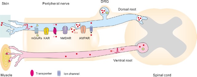
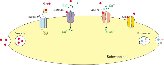
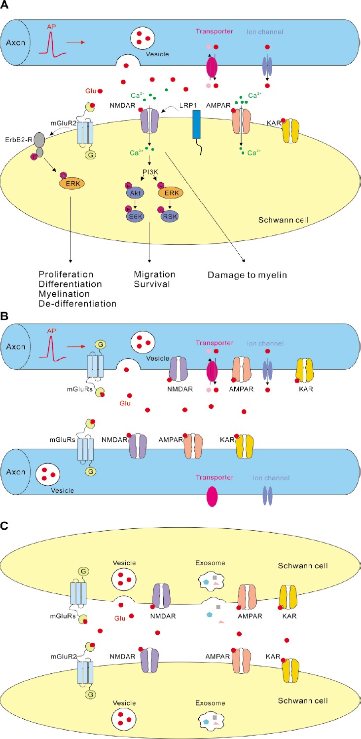
In the peripheral nervous system, the vast majority of axons are accommodated within the fibre bundles that constitute the peripheral nerves. Axons within the nerves are in close contact with myelinating glia, the Schwann cells that are ideally placed to respond to, and possibly shape, axonal activity. The mechanisms of intercellular communication in the peripheral nerves may involve direct contact between the cells, as well as signalling via diffusible substances. Neurotransmitter glutamate has been proposed as a candidate extracellular molecule mediating the cross-talk between cells in the peripheral nerves. Two types of experimental findings support this idea: first, glutamate has been detected in the nerves and can be released upon electrical or chemical stimulation of the nerves; second, axons and Schwann cells in the peripheral nerves express glutamate receptors. Yet, the studies providing direct experimental evidence that intercellular glutamatergic signalling takes place in the peripheral nerves during physiological or pathological conditions are largely missing. Remarkably, in the central nervous system, axons and myelinating glia are involved in glutamatergic signalling. This signalling occurs via different mechanisms, the most intriguing of which is fast synaptic communication between axons and oligodendrocyte precursor cells. Glutamate receptors and/or synaptic axon-glia signalling are involved in regulation of proliferation, migration, and differentiation of oligodendrocyte precursor cells, survival of oligodendrocytes, and re-myelination of axons after damage. Does synaptic signalling exist between axons and Schwann cells in the peripheral nerves? What is the functional role of glutamate receptors in the peripheral nerves? Is activation of glutamate receptors in the nerves beneficial or harmful during diseases? In this review, we summarise the limited information regarding glutamate release and glutamate receptors in the peripheral nerves and speculate about possible mechanisms of glutamatergic signalling in the nerves. We highlight the necessity of further research on this topic because it should help to understand the mechanisms of peripheral nervous system development and nerve regeneration during diseases.
Keywords: AMPA receptors; NMDA receptors; PNS; Schwann cells; axons; glutamate; metabotropic glutamate receptors; myelination; nerve injury; peripheral nervous system; synaptic signalling.
Conflict of interest statement
None
Figures
References
-
- Bacich DJ, Wozniak KM, Lu XC, O’Keefe DS, Callizot N, Heston WD, Slusher BS. Mice lacking glutamate carboxypeptidase II are protected from peripheral neuropathy and ischemic brain injury. J Neurochem. 2005;95:314–323. - PubMed
-
- Battaglia G, Rustioni A. Coexistence of glutamate and substance P in dorsal root ganglion neurons of the rat and monkey. J Comp Neurol. 1988;277:302–312. - PubMed
-
- Bhave G, Karim F, Carlton SM, Gereau RWt Peripheral group I metabotropic glutamate receptors modulate nociception in mice. Nat Neurosci. 2001;4:417–423. - PubMed
Publication types
LinkOut - more resources
Full Text Sources
